
Dr Matthew Seager amp Dr Miles Walkden BSGARBSUR Interesting Cases 2019 m atthewseager1nhsnet Clinical information 25 male 2 week referral by GP due to 34 cm lump superior to the left testicle ID: 930265
Download Presentation The PPT/PDF document "A case that left us wondering..." is the property of its rightful owner. Permission is granted to download and print the materials on this web site for personal, non-commercial use only, and to display it on your personal computer provided you do not modify the materials and that you retain all copyright notices contained in the materials. By downloading content from our website, you accept the terms of this agreement.


Slide1
A case that left us wondering...
Dr Matthew Seager & Dr Miles WalkdenBSGAR-BSUR Interesting Cases 2019matthew.seager1@nhs.net
Slide2Clinical information25 male
2 week referral by GP due to “3-4 cm” lump superior to the left testicleFelt to be epididymal cyst, but could be testicular in origin O/E
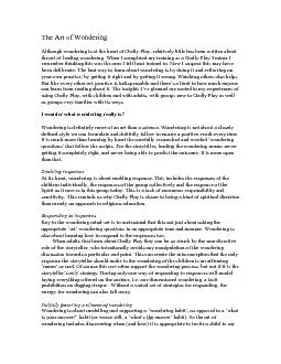
Slide3Longitudinal section ultrasound image - left paratesticular region. * - epididymis thought to be separate.
*
Slide4Longitudinal section ultrasound image - left paratesticular region with colour Doppler
Slide5Transverse section ultrasound image
– left paratesticular region with colour Doppler
Slide6Differential diagnosis
Epididymal cyst Solid paratesticular lesions:
Adenomatoid tumour
Spermatic cord lipoma
Leiomyoma
Haemangioma
Sarcoma
MDT decision: given vascularity -> surgical biopsy +/- excision
✗
- not cystic
? -
extremely rare, but is vascular
?
- usually epididymal - tail
?
-
long history of lump
?
-
atypical appearance
?
-
not typically so vascular
Slide7Final diagnosisLesion separate from epididymis at surgery
Closely associated with testicular vessels so not removed3 x biopsies obtained:Ectopic splenic tissueGiven the location - final diagnosis of splenogonadal fusion
Slide8White pulp
Red pulp
Slide9Discussion
Splenogonadal fusion:M:F 15:1, nearly all leftClose proximity of developing gonad and spleen in embryonic weeks 5-8Continuous or discontinuousContinuous variety – 1/3 congenital abnormalities e.g. cryptorchidism, limb defects
Fusion with mesonephric derivatives rarely
Slide10Discussion
Presentation40% scrotal swelling20% inguinal hernia18% autopsy diagnosis15% cryptorchidism7% other
ImagingSlight reduced reflectivity relative to testisMay be difficult to separate from testis
–
DD for testicular Ca
Central vascular pattern branching to periphery c.f. disorganised malignant
Difficult to pre-operatively suspect but
99m
Tc sulphur colloid diagnostic
Varma et al.
Marko et al.
Slide11Discussion
Longitudinal section ultrasound image
Slide12Discussion
ManagementAvoid unnecessary orchidectomy!No evidence increased malignancy
✗
Slide13Key learning points
Rare conditionConsider splenogonadal fusion with a paratesticular mass and congenital anomaliesAwareness may allow appropriate pre-operative workup to avoid orchidectomyImportant to be aware of more common causes for solid paratesticular lesions
Slide14References
Putschar WGJ, Manion WC. Splenic-gonadal fusion. Am J Pathol. 1956;32: 15–33. Carragher AM. One hundred years of splenogonadal fusion. Urology.
1990;35: 471–75. Varma et al.
Sonographic
and CT features of splenogonadal fusion.
Pediatr
Radiol
. 2007;37(9): 916-9.
Marko et al. Testicular seminoma and its mimics.
Radiographics
. 2017;37(4): 1085-98.
Stewart VR,
et al.
Splenogonadal fusion. B-mode and color Doppler sonographic appearances. J Ultrasound Med. 2004;23: 1087-90.