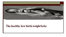
/
The  healthy low birth weight baby The  healthy low birth weight baby

The healthy low birth weight baby - PowerPoint Presentation

BabyDoll
BabyDoll . @BabyDoll
Follow
342 views
Uploaded On 2022-08-01

The healthy low birth weight baby - PPT Presentation

Classification of babies by gestation and weight Definitions of gestational age disregard any considerations of birth weight and likewise definitions of LBW are based upon weight alone and do not consider the gestational age of the baby ID: 931779

babies baby growth gestational baby babies gestational growth age birth weight preterm small glucose skin fetal head gestation term

Share:

Link:

Embed:

Download Presentation from below link

Download Presentation The PPT/PDF document "The healthy low birth weight baby" is the property of its rightful owner. Permission is granted to download and print the materials on this web site for personal, non-commercial use only, and to display it on your personal computer provided you do not modify the materials and that you retain all copyright notices contained in the materials. By downloading content from our website, you accept the terms of this agreement.


Presentation Transcript

Slide1

The

healthy low birth weight baby

Slide2

Classification of babies by gestation and weightDefinitions of gestational age disregard any considerations of birth weight and likewise definitions of LBW are based upon weight alone and do not consider the gestational age of the baby.

Slide3

Gestational ageAccording to Smith (2012), babies should preferably be classified by gestational age, as this is a befer physiological measure compared to birth weight. A preterm baby is born before completion of the 37th gestational week (259 days), which is calculated from the first day of the mother's last menstrual period (LMP) and has no relevance to the baby's weight, length, head circumference, or indeed any other measurement of fetal or neonatal size.

Slide4

Smith (2012) further asserts that gestational age estimates by first- trimester ultrasonography are accurate within 4 days, so that the combination of fetal crown–rump length and menstrual history are now considered more accurate indices for estimating gestational age.

Slide5

Birth weightThe World Health Organization (WHO 1997a) recommend that babies who weigh <2500 g should be called low birth weight (LBW). As neonatal care has become more effective and babies are surviving at earlier gestations, new LBW categories are now recognized:• very low birth weight (VLBW) babies are those weighing below 1500 g at birth• extremely low birth weight (ELBW) babies are those who weigh below 1000 g at birth.

Slide6

It is the relationship between weight (for assessment of growth) and gestational age (for assessment of maturity) that is of great importance. This relationship can be seen plotted on centile charts (Fig. 30.1) to visually demonstrate that growth is appropriate, excessive or diminished for gestational age and that the baby is either preterm, term or post-term.

Slide7

Growth charts, however, should be derived from studies of local populations, because genetically derived growth differences exist between countries, cultures and lifestyles.

Slide8

FIG. 30.1 A centile chart, showing weight and gestation.

Slide9

Various presentations of LBW babies can be described as follows:1. Babies whose rate of intrauterine growth is normal at the moment of birth. They are small because labour began before the end of the 37th gestational week. These preterm babies are appropriately grown for their gestational age (AGA). Their weight is between the 90th and 10th centiles for their gestation age.

Slide10

2. Babies whose rate of intrauterine growth has slowed down and who are born at, or later than, term. These term or post-term babies are under-grown for gestational age and are consequently small for their gestational age (SGA). Their weight is below the 10th centile for their gestational age.

Slide11

3. Babies whose rate of intrauterine growth has slowed down and who are also born before term. These preterm babies are small by virtue of both their early birth and impaired intrauterine growth. They are both pre-term and SGA babies because their weight will be below the 10th centile for their reduced gestational age.

Slide12

Babies are considered large for their gestational age (LGA) when their weight exceeds the 90th centile. Consequently, it should be recognized that both term and preterm babies can fall into the category of AGA, SGA, or LGA (Fig. 30.2).

Slide13

Small for gestational age (SGA)Babies that are small for their gestational age are of a size that is smaller when compared to other babies. If a baby is under-grown and below the 10th centile for weight, historically there has been for some an automatic assumption that as a fetus the baby has experienced intrauterine growth restriction (IUGR). Wilkins-Haug and Heffner (2012) define IUGR as a rate of fetal growth that is less than the normal growth potential for a specific baby.

Slide14

However, this does not mean that all SGA babies are small as a result of IUGR. Some small babies are genetically small because they have small parents or grandparents and this familial factor determines their smallness. They are well, healthy babies who need to be treated accordingly.

Slide15

Centile charts can act only as guides. Trotter (2009) states that maternal characteristics, obstetric history and birth details in addition to the appearance and behaviour of the baby should determine what care is required.Should a baby be born at 36 weeks' gestation with a birth weight of 2100 g, which according to weight, is well below the 50th centile, this baby would not fall below the 10th centile line for weight, so should not be identified as SGA but may be under-grown.

Slide16

Similarly it should not be assumed that all infants of diabetic mothers (IDM) are macrosomic and only fall into the LGA category. Diabetes and obesity are conditions that deleteriously affect maternal circulation and perfusion, so some babies will suffer from IUGR and could be small for their gestational age.

Slide17

Types of intrauterine growth restriction (IUGR)There are two recognized types of IUGR. The causes and predisposing factors are seen as multi-factorial (Box 30.1)In the book

Slide18

1. IUGR that begins early in the first trimester caused by a combination of intrinsic and extrinsic factors, results in symmetrical fetal growth

Slide19

In this scenario, the fetus suffers significant interruption to hyperplastic cell division . As a result, the head circumference, length

and w

e

i

g

h

t

are

all

proportionately

reduced

for

gestational

age.

The

main

causes

are

referred

to

as

intrinsic

factors

that

operate

from

within

the

fetus

and

cause

symmetrical

g

ro

w

t

h

restriction,

o

ft

en

as

a

result

of

transplacental

infections

or

chromosomal/genetic

defects.

Slide20

In addition, the deleterious effects of maternal lifestyle where a poor quality diet may be in combination with smoking, drug and/or alcohol misuse, can impact on fetal growth and development. These examples are referred to as extrinsic factors that can act upon the fetal environment and contribute to congenital malformations that culminate in conditions such as fetal alcohol syndrome (FAS) or chronic hypoxia associated with maternal smoking.

Slide21

Affected babies suffer interruption to hyperplastic (new cell) division, therefore look small and do not have the potential for normal growth. Remember a small head equates to a small brain.

Slide22

2. IUGR that begins in the last trimester, caused by extrinsic factors, results in asymmetrical fetal growth

Slide23

This type of fetus has been growing normally then starts to experience interruption to hypertrophic cell growth.

This is influenced

by

e

x

t

r

i

n

s

i

c

factors

in

its

intrauterine

environment

that

cause

disruption

to

placental

perfusion

of

oxygen

and

nutrients.

Slide24

When serial ultrasound scans of head and abdominal circumference in addition to Doppler measurements indicate poor and disproportionate growth, the birth of many affected fetuses are expedited early, usually by elective caesarean section.The baby's head appears relatively large compared to the body (see Fig. 30.4); however, the head circumference is usually within normal parameters and brain growth is usually spared.

Slide25

The skull bones are within gestational norms for length and density but the anterior fontanelle may be larger than expected, owing to diminished bone formation.The abdomen appears sunken owing to shrinkage of the liver and spleen, which surrender their stores of glycogen and red blood cell mass respectively as the fetus adapts to the adverse conditions of the uterus.

Slide26

As subcutaneous fat is used as a source of glucose and ketones, the skin becomes loose, giving the baby a wizened, old appearance. Vernix caseosa is frequently reduced or absent as a result of diminished skin perfusion. In the absence of this protective covering, the skin is continuously exposed to amniotic fluid and its cells will begin to desquamate (shed) so that the skin appears pale, dry and coarse.

Slide27

If the baby is of a mature gestation and has passed meconium in utero, the skin may be stained with meconium. Fetal distress in labour and hypoglycaemia are more likely to be seen in this group of babies. Unless severely affected, these babies appear hyperactive and hungry, with a lusty cry.

Slide28

FIG. 30.4 Baby with asymmetrical growth restriction. Note the apparently large head compared with the undergrown body.

Slide29

 The preterm babyThe preterm baby is born before the end of the 37th gestational week, regardless of birth weight. Most of these babies are appropriately grown, some are SGA and a small number are LGA. The factors that play a role in the initiation of preterm labour are largely unknown and mainly overlay with factors that impair fetal growth.

Slide30

They are divided into those labours that commence spontaneously and those where a decision is made to terminate a viable pregnancy before term: referred to as elective causes (Box 30.2). In the book

Slide31

Characteristics of the preterm babyThe appearance of the preterm baby at birth will depend upon the gestational age. The following description will focus upon the baby born from 32 weeks' gestation. Preterm babies rarely grow large enough in utero to develop muscular flexion and fully adopt the fetal position. As a result their posture appears flattened, with hips abducted, knees and ankles flexed.

Slide32

Lissauer and Faranoff (2011) describe a generally hypotonic baby with a weak and feeble cry. The head is in proportion to the body, the skull bones are soft with large fontanelles and wide sutures. The chest is small and narrow and appears underdeveloped.The abdomen is prominent because the liver and spleen are large and abdominal muscle tone is poor (Fig. 30.5).

Slide33

FIG. 30.5 Healthy preterm baby born at 32 weeks' gestation. Note the presence of a nasogastric tube. The thermocouple of the servo-mode is taped to the skin of the baby's upper abdomen.

Slide34

The liver is large because it receives a good supply of oxygenated blood and is active in the production of red blood cells. The umbilicus appears low in the abdomen because linear growth is cephalo-caudal, being more apparent nearer to the head than rump, by virtue of fetal circulation oxygenation.Subcutaneous fat is laid down from 28 weeks' gestation, therefore its presence and amount will affect the redness and transparency of the skin.

Slide35

Vernix caseosa is abundant in the last trimester and tends to accumulate at sites of dense lanugo growth, such as the face, ears, shoulders and sacral region, protecting the skin from amniotic fluid maceration. The ear pinna is flat with little curve, the eyes bulge and the orbital ridges are prominent. The nipple areola is poorly developed and barely visible. The cord is white, fleshy and glistening.

Slide36

The plantar creases are absent before 36 weeks' gestation but soon begin to appear, as fluid loss occurs through the skin. In girls the labia majora fail to cover the labia minora and in boys the testes descend into the scrotal sac at about the 37th gestational week.

Slide37

cephalocaudal development :  proceeding or occurring in the long axis of the body especially in the direction from head to tail 

Slide38

Hypo glycaemiaThe term hypo glycaemia refers to a low blood glucose concentration and is usually a feature of failure to adapt from the fetal state of continuous trans placental glucose consumption to the extra uterine pattern of intermittent milk supply ( WHO 1997b).

Slide39

Within the first hour of life the blood glucose levels fall, which triggers the pancreas to stimulate the alpha cells of the Islets of Langerhans to produce glucagon, with the consequential effect of releasing glucose from glycogen stores in the liver to maintain the blood glucose levels within safe limits.

Slide40

However, it is generally questioned whether LBW babies are as effective in this metabolism compared to appropriately grown term babies and some caution is recommended (WHO 1997b). Asymmetrical SGA babies may have greater brain-to-body mass with a tendency towards polycythemia, which increases their energy demands, and since both the brain and the red blood cells are obligatory glucose users, these factors can increase glucose requirements.

Slide41

Glycogen storage is initiated at the beginning of the third trimester of pregnancy but may be incomplete as a result of preterm birth or, in the asymmetrical SGA baby, may have been drawn upon before birth.

Slide42

Hypo glycaemia in healthy LBW babies is more likely to occur in conditions where they become cold or where the initiation of early feeding (within the first hour) is delayed. However, hypo glycaemia is associated with mild to moderate perinatal asphyxia and maternal history of beta-blocker use (e.g. labetolol) as it causes hyperinsulinism and interferes with glycogenolysis.

Slide43

The midwife should consider that there may be some underlying medical condition that may call for more thorough investigation.The signs of hypoglycaemia are varied and Wilker (2012) acknowledges that hypoglycaemia can present with no or few clinical signs. The clinical picture of tremor and irritability may occasionally lead to convulsion and decreased consciousness.

Slide44

A high- pitched cry, hypotonia, unexplained apnoea and bradycardia with central cyanosis are also recognized as serious signs of deterioration in the baby's health and need referral to a medical practitioner.

Slide45

The aim of management is to maintain the true blood glucose level above 2.6 mmol/l, which is considered to be the lowest level of normal in the first few days of life (WHO 1997b; Lissauer and Fanaroff 2011).Healthy LBW babies who show no clinical signs of hypoglycaemia, are demanding and taking nutritive feeds on a regular basis and maintaining their body temperature, do not need screening for hypoglycaemia.

Slide46

The emphasis of care is placed upon the concept of adequate feeding and the cornerstone of success is the midwife's ability to assess whether the baby is feeding sufficiently well to meet energy requirements. The preterm baby may be sleepy and attempts to take the first feed may reflect its gestational age.

Slide47

Midwives should be guided by the local policies within their employing organization regarding use of reagent strips to assess for hypoglycaemia, but prior to the baby's second feed is the best time to ascertain whether the first feed was effective in maintaining the capillary blood glucose level above 2 mmol/l. If a baby, despite being fed, presents with clinical signs of hypoglycaemia, a venous sample should be taken by the medical practitioner to assess the true blood glucose level which should be dispatched to the laboratory.

Slide48

A true blood glucose level that remains <2.6 mmol/dl, despite the baby's further attempts to feed by breast or take colostrum by cup, may warrant transfer to the NICU, because glucose by intravenous bolus may be necessary to correct the metabolic disturbance.

Slide49

Healthy mature SGA babies with an asymmetrical growth pattern will usually breastfeed within the first 30–60 minutes of birth and will demand feeds every 2–3 hours thereaher. For the majority of LBW babies, hypoglycaemia is relatively short-lived and limited to the first 48 hours following birth.

Slide50

FeedingAccording to Jones and Spencer (2008) both preterm and SGA babies benefit from human milk because it contains long chain polyunsaturated omega-3 fatty acids, which are thought to be essential for the myelination of neural membranes and for retinal development.Preterm breast milk has a higher concentration of lipids, protein, sodium, calcium and immunoglobulin's, alongside lipases and enzymes that improve digestion and absorption.

Slide51

The uniqueness of the mother's milk for her own baby cannot be overstated but she needs to understand what her baby may be able to achieve related to the stage of their development, which is based upon the combined influences of their gestational age at birth and their neonatal age.

Slide52

For a baby to feed for nutritive purposes, the coordination of breathing with suck and swallow reflexes reflects neuro behavioral maturation and organization, which is thought to occur between 32 and 36 weeks' gestation. Blackburn (2007) argues that preterm babies are limited in their ability to suck because they lack cheek pads, which leads to a weaker suck, coupled with weak musculature and flexor control, which are important for firm lip and jaw closure.

Slide53

The preterm baby's head is very heavy for the weak musculature of the neck and would, if not supported, result in considerable head lag, so correct positioning and attachment to the breast can be made much more difficult to achieve. Poor head alignment can result in airway collapse, which may lead to apnea and bradycardia, therefore support from the midwife is essential when initiating breastfeeding.

Slide54

If the baby requires feeding via a nasogastric tube, it is now common practice for parents to feed their own baby. Tube feeding has the advantage that the tube can be left in situ during a cup or breastfeed and has been shown to eliminate the need to introduce bottles into a breastfeeding regimen.

Slide55

However, babies are preferential nose breathers and the presence of a nasogastric tube will inevitably take up part of their available airway.Flint et al (2007) argue that the prolonged use of nasogastric tubes has been associated with delay in the development of a baby's sucking and swallowing reflexes simply because the mouth is bypassed.

Slide56

For these reasons, cup feeding has been used in addition or as an alternative to tube feeding, in order to provide the baby with a positive oral experience, to stimulate saliva and lingual lipases to aid digestion and to accelerate the transition from naso/oro-gastric feeding to breastfeeding. Oral gastric tubes have been associated with vagal stimulation and have resulted in bradycardia and apnoea.

Slide57

licking and lapping, are well established before sucking and swallowing, and when babies are given the opportunity it is not unusual to see them as early as 28 and 29 weeks licking milk that has been expressed onto the nipple by their mother. Thus, babies between 30 and 32 weeks' gestation can be given expressed breast milk (EBM) by cup. The baby uses less energy to take its feed by cup compared to bottle, which supports their general wellbeing and homeostasis.

Slide58

 Optimizing the care environment for the healthy LBW babyHandling and touchKangaroo care (KC) is used to promote closeness between a baby and mother and involves placing the nappy-clad baby upright between the maternal breasts for skin-to- skin contact (Fig. 30.6).

Slide59

Kangaroo care

Slide60

The LBW baby can remain beneath the mother's clothing for varying periods of time that suit the mother. Some mothers may have repeated contacts throughout the day, others may prefer specific periods around which they plan their daily activities. There are no rules or time limitations applied, but contact should be reviewed if there are any clinical signs of neonatal distress.

Slide61

Hake-Brooks and Anderson (2008) found that preterm babies of 32–36 weeks' gestation who had unlimited skin-to- skin contact, breast fed for longer compared to those who had traditional nursery care. Conde-Agudelo and Belizan (2009) support this view and also consider that the baby remained more physiologically stable, with less reported incidence of infection

Slide62

 Sleeping position