
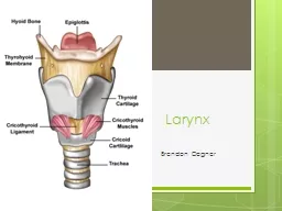
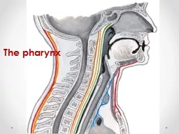
Anatomy of the larynx The larynx is made of Hyoid bone U shaped near C3 level 9 cartilages 3 paired and 3 single cartilages Ligaments and membranes that connects the cartilage to give it stability ID: 932536
Download Presentation The PPT/PDF document "Anatomy of the larynx and benign lesions..." is the property of its rightful owner. Permission is granted to download and print the materials on this web site for personal, non-commercial use only, and to display it on your personal computer provided you do not modify the materials and that you retain all copyright notices contained in the materials. By downloading content from our website, you accept the terms of this agreement.


Slide1
Anatomy of the larynx and benign lesions of the larynx
Slide2Anatomy of the larynx
The larynx is made of:
Hyoid bone : U shaped, near C3 level
9 cartilages: 3 paired and 3 single cartilagesLigaments and membranes that connects the cartilage to give it stability2 set of muscles: intrinsic muscles the control the tension & orientation of the vocal cordsExtrinsic muscles that adjust the position of larynx during swallowingRespiratory mucosa: covers the interior surface of the larynx which is continuous above with the pharynx and below with trachea
Slide3Cartilages
Single cartilages
1. Thyroid cartilage:
The most prominent & the largest laryngeal cartilage2. cricoid cartilage:Located at C6It has a signet ring shapeThe only cartilage that forms a complete Ring in the respiratory system3. Epiglottis:
Thin Leaf likeCovered with pale mucous membraneAt the level of C5
Paired cartilages
1. arytenoid cartilage:
The
chief
moving part of the larynx
Articulates with
posterior
superior
surface of
cricoid
cartilage
2.
corniculate
cartilage:
3.
cuniform
cartilage:
Slide4Slide5True vocal folds
Epithelium:
stratified
nonkertinized squamous epitheliumlamina propria: A highly specialized lamina propria separates the epithelium from underlying muscle.Vocalis
muscle (Medial part of the Thyroarytenoid muscle)
Slide6The larynx is subdivided into
From
To
Supraglottic
area
(
Epiglottis, Arytenoids cartilage,
Corniculate
cartilage,
Conieform
cartilage, Aryepiglottic fold,
Interartenoid
notch, False vocal cord)
Epiglottissuperior border of the true vocal cordGlottis area (It is the narrowest part of the larynx in adults)Vertical plan.5-1cm below the free border of true VCSubglottic area (It is the narrowest part of the larynx in pediatrics).5 -1 cm below the free border of true V.CInferior border of the cricoids cartilage
Slide7Nerves
2 branches of
vagus
nerve:Superior laryngeal nerve:Supplies the cricothyroid muscleRecurrent laryngeal nerve:Gives motor innervations to all ipsilateral intrinsic laryngeal muscles except cricothyroid (superior laryngeal)
Slide8HOARSENESS IN VOICE
General term which describe
abnormal voice
changeHaving difficulty in producing the sound when trying to speakChange in a pitch and quality of the voiceThe voice may sound weak, very breathy, scratchy or husky
Slide9PHYSICAL EXAMINATION
RHINOLOGIC
&
OTOLOGIC EXAMINATION.NECK LYMPH NODE EXAMINATION.VISUALIZATION OF THE LARYNX BY:INDIRECT LARYNGOSCOPY.FIBEROPTIC NASOPHARYNGOSCOPY.
RIGID LARYNGOSCOPY.
Slide10INDIRECT LARYNGOSCOPY
Slide11Flexible Fiberoptic laryngoscopy
Slide12NORMAL LARYNGEAL STRUCTURES
Slide13Causes of Hoarseness of voice
Congenital
: laryngeal
web, cyst, laryngo
cele
Paralysis
: paralysis of
recurrent
laryngeal
nerve,
superior laryngeal
or both
Inflammation
: acute & chronic
laryngitis, laryngo-tracheo-bronchitis, diptheria, acute epiglottitisNeoplastic: vocal cord polyps, nodules, granuloma, cysts, laryngeal carcinoma, leukoplakia.Neuromuscular: vocal cord palsy, spasmodic dysphonia, movement disorder, Parkinson disease, CVA.Miscellaneous: vocal abuse, vocal cord atrophy, vocal cord scarring, hypothyroidism, Reinke’s edema, GERD, postnasal drip.
Slide14Infectious
type:
Follows URTI
Often viral in originBacterial will act as superadded infection; strepcoccus, H.influenza,
haemolytic strepcoccus & staph. Aureus.
Non infectious
type:
Vocal
abuse
Allergy
Smoking
/
alcohol
Thermal
/
chemical burn to larynxLaryngeal traumaAcute Laryngitis
Slide15Presentation
Aphonia
/ dysphonia
Cough: dry, painful & irritatingStridor: rare but potentially seriousPain throat: after talking
Examination:Indirect laryngoscopy
, shows
Red
swollen larynx
Sometimes, present
stringy mucus
between cords
Treatment
:
Vocal
rest
Avoidance of smoking & alcohol
Slide16Predisposing causes
:
Alcohol
Habitual shouting / faulty voice production Laryngeal muscle imbalance dysphonia Voice: hoarse & fatigue easily
Continues to smoke turn into carcinoma
Treatment
:
Voices should be
rested
Treat upper airway
sepsis
Steam inhalation
Chronic Laryngitis
Slide17Benign lesions of the larynx:
Vocal cord nodules
Singer’s nodule
Usually bilateral (rarely unilateral)small swellings (less than 3 mm in diameter)Location: the junction between the anterior 1/3 and the posterior 2/3 of the whole vocal cordTreatment: speech therapy is the mainstay, microlaryngoscopy and excision in refractory cases
Slide18Benign lesions of the larynx:
Vocal cord polyp
A true vocal polyp is a benign swelling of greater than 3 mm that arises from the free edge of the vocal fold
Polyps can shrink spontaneously or even be coughed up. Localized edema of the Reinke’s spaceMost need surgical removal
Slide19Leukoplakia (pre-malignant)
Localized form of
epithelial hyperplasia
Involving upper surface of one or both vocal cordAppears as white plaque or warty growth on the cord without affecting its mobilityTreatment: stripping of vocal cord & subjecting the tissue to histology for any malignant change
Slide20Vocal cord mobility disorders
Vocal cord paralysis:
Iatrogenic Injury (
most common cause) surgery: Thyroidectomy (the most common surgery )ant cervical fusionesophageal surgerycarotid endartectomy Mediastinal surgeryNeoplastic : Bronchial 50%:; Nasopharyngeal ca 20%; Esophageal 20 %; Thyroid 10%; lymphoma
Idiopathic (50% of the cases): usually self-limiting (take up to 12 months to resolve)Trauma
Neurological disease:
Infectious
:Lyme
disease; Syphilis;
EBV
; Tuberculosis;
Viral
Systemic Diseases
: Sarcoidosis;
D.M
Toxins: lead ; arsenic; quinine; Streptomyocin
Slide21Laryngomalacia
Most common cause of
stridor
in infants (60% of all laryngeal problem)Occur due to floppy supra-glottic tissueSymptoms: Inspiratory stridor that starts within 6 weeks of lifeExacerbating factors:Sleeping
Lying supineURTIRelieving factor:
lying prone
when the child is active
Diagnosis: Flexible
Fiberoptic
laryngoscopy
Treatment:
90%
will have their symptoms resolves within by
12 months
of ageOnly 10% will need surgical intervention
Slide22Acute laryngeal infections in Children
Acute epiglottitis/
supraglottitis
Acute
laryngotracheobronchitis
(croup)
Micro-organism
Bacteria: H. influenza type B
Viral: para-influenza type 1& 2
location
epiglottis
glottis and the subglottis
Onset
Short duration (hours)
Gradual (days)Age3-5 years6 months-3 yearsVoiceMuffled voice (not HOV)HOV symptoms
High grade fever
odynophagia+ dysphagia+ saliva drooling
sitting upright
Low grade Fever
Barking cough
Inspiratory stridor (early)
Xray
Thumb sign
Steeple sign
Treatment
Airway secured (priority)
antibiotics (corner stone)ex: cefuroxime
Steroid
Humidified O2
Nebulized epinephrine
Corticosteroid (single dose):
Croup
is the most common airway
obstructive infection
in children
Epiglottitis
Is inflammation of the
loosely
attached mucosa
Slide23Stridor
is an abnormal,
high-pitched
sound produced by turbulent airflow through a partially obstructed airway at the level of the supraglottis, glottis, subglottis and/or trachea.It should be differentiated from stertor
, which is a lower-pitched, snoring-type sound generated at the level of the
nasopharynx
,
oropharynx
& occasionally
supraglottis
.
Stridor is a
symptom
,
not a diagnosis
or disease, and the underlying cause must be determined.
Slide24Stridor depending on its timing in the respiratory cycle may be:
Inspiratory
stridor suggests a laryngeal obstruction.Expiratory stridor implies bronchial obstructionBiphasic
stridor suggests a tracheal (subglottic
or
glottic
anomaly).
Slide25Causes of acute stridor
Laryngo-tracheobronchitis (croup)
Aspiration of foreign body (
eg. peanut, coin, toys...) a history of Choking & coughing may precedes the development on RD symptomTracheitis, bacterial cause is most common in children <3 y, mainly staph aureus, viral influenza.
Retropharyngeal abscess is a complication of bacterial pharyngitis, observed in children <6 y.
Peritonsillar abscess, an infection in the potential space between superior constrictor muscle and tonsils.
Spasmodic
croup
Epiglottitis, which is a medical
emergency,most
commonly in children
2-7 y
.
Allergic
reaction within 30 min of adverse exposure
Slide26Causes of chronic stridor
Laryngomalacia
Vocal cord dysfunction
Subglottic stenosisLaryngeal websLaryngeal cystLaryngeal hamengiomas
TracheomalaciaLaryngeal papilloma