
Authors Curado S X K niggendorf V F K niggendorf S L J unior I R M Ugarte M F S INTRODUCTION CASE REPORT DISCUSSION REFERENCES Pathological myopia was originally described as myopia associated ID: 930674
Download Presentation The PPT/PDF document "IMPORTANCE OF OCTA IN HIGH MYOPIC PATIEN..." is the property of its rightful owner. Permission is granted to download and print the materials on this web site for personal, non-commercial use only, and to display it on your personal computer provided you do not modify the materials and that you retain all copyright notices contained in the materials. By downloading content from our website, you accept the terms of this agreement.


Slide1
IMPORTANCE OF OCTA IN HIGH MYOPIC PATIENTS WITH MACULA HEMORRHAGE
Authors
: Curado, S X; Kniggendorf, V F; Kniggendorf S L; Junior, I R M; Ugarte, M F S
INTRODUCTION
CASE REPORT
DISCUSSION
REFERENCES
Pathological myopia was originally described as myopia, associated with degenerative changes of the sclera, choroid and retinal pigment, epithelium.1,2 Choroidal neovascularization (CNV) is an important cause of visual impairment in this group. Fundus biomicroscopy, fluorescein angiography (AF), optical coherence tomography (OCT) are the recommended ancillary exams for CNV diagnosis in myopia. OCTA is a non-invasive diagnostic method that provides images of the retinal and choroidal circulation.3 OCTA could help to evaluate the presence and morphology of myopic CNV, which appears as a large hyperreflective vascular anastomotic network.6Two types of subretinal hemorrhage have been reported in myopia, those with and without choroidal neovascularization (CNV).Clinically, retinal changes, such lacker cracks, subretinalneovascular membranes and hemorrhages, are common in high myopia.4 In these cases hemorrhage could be absorved in a few months with a good prognosis, unless the bleeding recursor occurs a development of atrophic scars or degeneration of retina and choroid.5,6. Approximately 5 to 11% of high myopic patients will develop myopic CNV, and bilateral disease will develop in 35% of those patients within 8 years. Age at onset is an risk factor, with patients aged under 40 years with better prognosis.7 The treatment of myopic CNV with anti-VEGF is of fundamental importance and left untreated
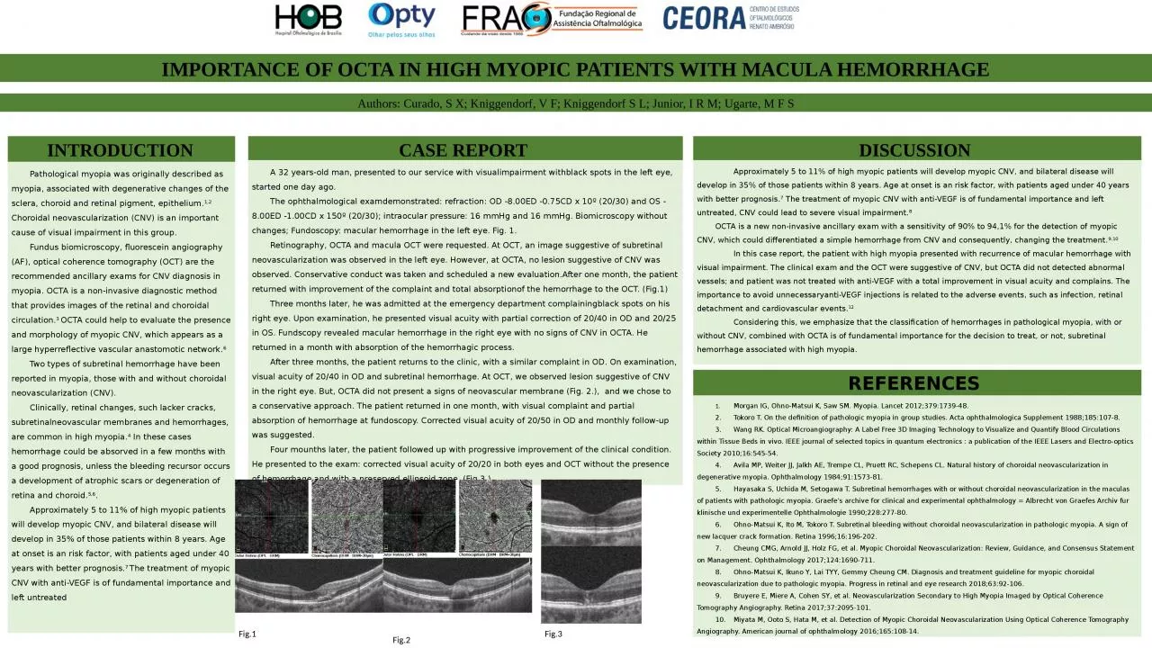
A 32 years-old man, presented to our service with visualimpairment withblack spots in the left eye, started one day ago.The ophthalmological examdemonstrated: refraction: OD -8.00ED -0.75CD x 10º (20/30) and OS -8.00ED -1.00CD x 150º (20/30); intraocular pressure: 16 mmHg and 16 mmHg. Biomicroscopy without changes; Fundoscopy: macular hemorrhage in the left eye. Fig. 1.Retinography, OCTA and macula OCT were requested. At OCT, an image suggestive of subretinal neovascularization was observed in the left eye. However, at OCTA, no lesion suggestive of CNV was observed. Conservative conduct was taken and scheduled a new evaluation.After one month, the patient returned with improvement of the complaint and total absorptionof the hemorrhage to the OCT. (Fig.1)Three months later, he was admitted at the emergency department complainingblack spots on his right eye. Upon examination, he presented visual acuity with partial correction of 20/40 in OD and 20/25 in OS. Fundscopy revealed macular hemorrhage in the right eye with no signs of CNV in OCTA. He returned in a month with absorption of the hemorrhagic process. After three months, the patient returns to the clinic, with a similar complaint in OD. On examination, visual acuity of 20/40 in OD and subretinal hemorrhage. At OCT, we observed lesion suggestive of CNV in the right eye. But, OCTA did not present a signs of neovascular membrane (Fig. 2.), and we chose to a conservative approach. The patient returned in one month, with visual complaint and partial absorption of hemorrhage at fundoscopy. Corrected visual acuity of 20/50 in OD and monthly follow-up was suggested. Four mounths later, the patient followed up with progressive improvement of the clinical condition. He presented to the exam: corrected visual acuity of 20/20 in both eyes and OCT without the presence of hemorrhage and with a preserved ellipsoid zone. (Fig 3.)
Approximately 5 to 11% of high myopic patients will develop myopic CNV, and bilateral disease will develop in 35% of those patients within 8 years. Age at onset is an risk factor, with patients aged under 40 years with better prognosis.7 The treatment of myopic CNV with anti-VEGF is of fundamental importance and left untreated, CNV could lead to severe visual impairment.8OCTA is a new non-invasive ancillary exam with a sensitivity of 90% to 94,1% for the detection of myopic CNV, which could differentiated a simple hemorrhage from CNV and consequently, changing the treatment.9,10 In this case report, the patient with high myopia presented with recurrence of macular hemorrhage with visual impairment. The clinical exam and the OCT were suggestive of CNV, but OCTA did not detected abnormal vessels; and patient was not treated with anti-VEGF with a total improvement in visual acuity and complains. The importance to avoid unnecessaryanti-VEGF injections is related to the adverse events, such as infection, retinal detachment and cardiovascular events.12 Considering this, we emphasize that the classification of hemorrhages in pathological myopia, with or without CNV, combined with OCTA is of fundamental importance for the decision to treat, or not, subretinal hemorrhage associated with high myopia.
1. Morgan IG, Ohno-Matsui K, Saw SM. Myopia. Lancet 2012;379:1739-48.2. Tokoro T. On the definition of pathologic myopia in group studies. Acta ophthalmologica Supplement 1988;185:107-8.3. Wang RK. Optical Microangiography: A Label Free 3D Imaging Technology to Visualize and Quantify Blood Circulations within Tissue Beds in vivo. IEEE journal of selected topics in quantum electronics : a publication of the IEEE Lasers and Electro-optics Society 2010;16:545-54.4. Avila MP, Weiter JJ, Jalkh AE, Trempe CL, Pruett RC, Schepens CL. Natural history of choroidal neovascularization in degenerative myopia. Ophthalmology 1984;91:1573-81.5. Hayasaka S, Uchida M, Setogawa T. Subretinal hemorrhages with or without choroidal neovascularization in the maculas of patients with pathologic myopia. Graefe's archive for clinical and experimental ophthalmology = Albrecht von Graefes Archiv fur klinische und experimentelle Ophthalmologie 1990;228:277-80.6. Ohno-Matsui K, Ito M, Tokoro T. Subretinal bleeding without choroidal neovascularization in pathologic myopia. A sign of new lacquer crack formation. Retina 1996;16:196-202.7. Cheung CMG, Arnold JJ, Holz FG, et al. Myopic Choroidal Neovascularization: Review, Guidance, and Consensus Statement on Management. Ophthalmology 2017;124:1690-711.8. Ohno-Matsui K, Ikuno Y, Lai TYY, Gemmy Cheung CM. Diagnosis and treatment guideline for myopic choroidal neovascularization due to pathologic myopia. Progress in retinal and eye research 2018;63:92-106.9. Bruyere E, Miere A, Cohen SY, et al. Neovascularization Secondary to High Myopia Imaged by Optical Coherence Tomography Angiography. Retina 2017;37:2095-101.10. Miyata M, Ooto S, Hata M, et al. Detection of Myopic Choroidal Neovascularization Using Optical Coherence Tomography Angiography. American journal of ophthalmology 2016;165:108-14.
Fig.1
Fig.3
Fig.2