
Epithelium Lecture Objectives Describe the structural and functional relationship between epithelium and connective tissue and epithelium and basal lamina Describe the structural and functional features of epithelial intercellular junctions and specializations of the apical surface of epithel ID: 934000
Download Presentation The PPT/PDF document "Epithelium Kristine Krafts, M.D." is the property of its rightful owner. Permission is granted to download and print the materials on this web site for personal, non-commercial use only, and to display it on your personal computer provided you do not modify the materials and that you retain all copyright notices contained in the materials. By downloading content from our website, you accept the terms of this agreement.


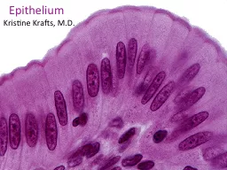
Slide1
Epithelium
Kristine Krafts, M.D.
Slide2Epithelium Lecture ObjectivesDescribe the structural and functional relationship between:epithelium and connective tissue, and
epithelium and basal lamina.Describe the structural and functional features of epithelial intercellular junctions and specializations of the apical surface of epithelial cells.
Slide3More Epithelium Lecture ObjectivesDescribe the structural and functional diversity of epithelium.
Describe the classification system of covering/lining epithelium.
Describe the histologic features of glandular epithelium
.
Slide4Epithelium Lecture OutlineFunction and types of epitheliumStructure of epitheliumTypes of covering/lining epithelium
Types of glandular epithelium
Slide5Epithelium Lecture OutlineFunction and types of epithelium
Slide6Functions of EpitheliumCovering of external surfaces Lining of internal surfaces Protection
AbsorptionSecretionSensation Contraction
Slide7Two Main Kinds of EpitheliumCovering and lining epithelium
Covers outer surfaces of body and lines internal body passagesGlandular epitheliumContains cells specialized for secretion
Slide8Unique Characteristics of EpitheliumBasal lamina anchors epithelium to underlying connective tissue.
Epithelial cells are very cohesive due to intercellular junctions.Epithelial cells
vary
a lot in shape and size.
Epithelial tissues are
avascular
.
Epithelial cells demonstrate
polarity
.
Slide9Epithelium Lecture OutlineFunction and types of epithelium
Structure of epitheliumBasement membraneConnections between cells
Specialized apical structures
Slide10Epithelium Lecture OutlineFunction and types of epithelium
Structure of epitheliumBasement membrane
Slide11Basement membrane
Epithelial cells
Slide12Basement
membrane =
Basal Lamina + Reticular Lamina
Basement membrane
Epithelium
Basal
lamina
Reticular
lamina
Slide13Basal
lamina
Reticular
lamina
Anchoring fibrils
(type VII collagen)
Anchoring plaques
(type IV collagen)
Reticular fibers
(type III collagen)
Epithelial cell
Type IV collagen and
perlecan
(a proteoglycan)
Laminin
(a glycoprotein)
lamina lucida
lamina densa
Composition of Basal Lamina and Reticular Lamina
Slide14Functions of Basal LaminaStructure: attaches epithelium to connective tissue Organization
: arranges plasma membrane proteins in the basal membraneFiltration: regulates movement of material between epithelium and connective tissue
Slide15Don’t make this mistake! “Basal lamina” and “basement membrane” are sometimes used interchangeably.
This is wrong, wrong, wrong!The basal lamina is part of the basement membrane. They are not the same thing.
Slide16Epithelium Lecture OutlineFunction and types of epithelium
Structure of epitheliumBasement membraneIntercellular junctions
Slide17Intercellular junctions connect epithelial cellsIntercellular junctions are present in most tissues but are especially numerous and prominent in epithelium.
Zonula occludens (tight junction)Zonula adherens (belt desmosome)Macula adherens (desmosome)Hemidesmosomes
Gap junction (nexus)
Slide18Intercellular Junctions
Slide19Intercellular junctions
(Zonula adherens)
(Zonula occludens)
Slide20Zona occludens (tight junction)
Most apical junction in epithelium
Form bands (zonula) that completely encircle each cell
Membranes of adjacent cells fuse to seal off the intercellular space
Slide21Zona occludens (tight junction)
Claudins and occludins are two families of proteins that form a seal to prevent flow of materials between epithelial cells
More zona occludens = tighter seal
Slide22Slide23Zonula adherens (belt desmosome)
Form bands that completely encircle each cell
Cadherin and catenin proteins provide adhesion between adjacent cells
Actin filaments in cytoplasm insert into attachment plaques
Slide24Macula adherens (spot desmosome)
Spot adhesion between cells
Desmosomes on adjacent cells line up
Cadherins present in intercellular space
Slide25Macula adherens (spot desmosome)Cytokeratin intermediate filaments insert into attachment plaques containing desmoplakin
and plakoglobinSuper strong attachment points between cells
The more desmosomes, the more tightly the epithelial cells are attached
Slide26Macula adherens (spot desmosome)
Slide27Pemphigus vulgaris
A blistering disease in which patients make autoantibodies to desmoglein proteins
Epithelial cell connections (spot desmosomes) loosen, causing fluid accumulation and superficial blisters
Slide28Cytokeratin
i
ntermediate
filaments
Cytoplasmic
attachment
plaques
Plasma membranes
Macula adherens (spot desmosome)
Slide29Spot desmosomes in stratum spinosum of skin
Slide30Hemidesmosomes
attach epithelial cells to basal lamina.
Integrins attach the basal portion of the cell to the basal lamina.
Hemidesmosomes
Slide31Keratin intermediate filaments in epithelial cell
Hemidesmosomes
Attachment plaque
Reticular lamina
Slide32Bullous pemphigoid
A blistering disease in which patients make autoantibodies to “bullous pemphigoid antigen” in hemidesmosome attachment plaques
Epithelial cells detach from basal lamina, causing fluid accumulation and blister formation
Slide33Junctional complex of intercellular junctionsZO: zonula occludensZA: zonula adherens
D: desmosomeIn some types of epithelia (simple columnar of digestive tract) junctions occur in this order
Slide34Gap (communicating) junctionOccur almost anywhere along lateral surfaces of epithelial cells and also in other cells, such as cardiac muscle cells
Connexons are protein channels with central pores connecting plasma membranes Allow ions and other small molecules to pass through to adjacent cells to facilitate communication
Slide35name
tight
junction
adherens
junction
desmosome
junction
gap
junction
hemidesmosome
junction
Slide36Epithelium Lecture OutlineFunction and types of epithelium
Structure of epitheliumBasement membrane
Intercellular junctions
Specialized apical structures
Slide37Microvilli
Microvilli + cell coat (or glyocalyx) = brush border or striated border
Purpose of microvilli: increase surface area for absorption or secretion
Slide38Microvillus
Tight junctions
Belt desmosome
Spot desmosome
Gap junction
Intermediate
filament
Hemidesmosome
Basal lamina
Slide39Microvilli
Microvilli have a
central core of
actin
microfilaments
Microvilli don’t wave back and forth like cilia.
Slide40Stereocilia
Stereocilia
are long,
non-motile microvilli found in parts of the male reproductive system
Stereocilia
Spermatozoa
Slide41Cilia
Cilia
are much longer and wider than microvilli.
They move back and forth to propel fluid along the epithelial surface.
Cilia on respiratory epithelial cells
Slide42Cilia
Cilia contain microtubules in a 9 + 2 configuration called an “
axoneme
”
2 central microtubules surrounded by 9 pairs of microtubules
Cilia insert into
basal bodies
with 9 triplets of microtubules
Slide43Epithelium Lecture OutlineFunction and types of epithelium
Structure of epitheliumTypes of covering/lining epithelium
Slide44How is covering/lining epithelium categorized?Shape of superficial cells
Number of cell layersPresence of specialized structures
Slide45How is covering/lining epithelium categorized?Shape of superficial cells
Squamous: width > height (flattened)Cuboidal: width = height (square, round)Columnar: width < height (tall
and
slender
)
Slide46Epithelial cells have different shapes
squamous cells are flat
cuboidal cells are cute and boxy
c
olumnar cells are tall and regal
Slide47How is covering/lining epithelium categorized?Shape of superficial cells
Number of cell layersSimple: one layer of cellsStratified: two or more layers of cells
Pseudostratified: all cells contact basal lamina,
but not all cells reach lumen
Slide48Simple epithelium: one layer of cells
Simple squamous epithelium
Simple cuboidal epithelium
Simple ciliated columnar epithelium
Slide49Endothelium is simple squamous epithelium.
It lines blood and lymphatic vessels.
Simple Squamous Epithelium
Flattened squamous cells
in a single layer
Slide50Mesothelium is simple squamous epithelium.
It lines serous cavities (pleura, pericardium, peritoneum).
Simple Squamous Epithelium
Flattened squamous cells
in a single layer
Slide51Duct linings often have simple cuboidal epithelium,
like this smallish duct in the pancreas.
Simple Cuboidal Epithelium
Cuboidal cells
in one cute layer
Slide52Stratified and pseudostratified epithelium
Stratified squamous epithelium
Pseudostratified columnar epithelium
Slide53Stratified Squamous Epithelium
Several layers of squamous epithelial cells
Mucous membranes are composed of stratified squamous epithelium.
Slide54Some ducts are lined by stratified cuboidal epithelium,
like this larger duct in the pancreas.
Stratified Cuboidal Epithelium
Cuboidal cells
in a few layers
Slide55How is covering/lining epithelium categorized?Shape of superficial cells
Number of cell layersPresence of specialized structuresCilia
Microvilli
Keratin
Slide56Pseudostratified Ciliated Columnar Epithelium
g
oblet cell
cilia
Respiratory epithelium is pseudostratified columnar,
with goblet cells and ciliated cells.
Slide57Simple Columnar Epithelium
The epithelium of the small intestine is simple columnar,
with goblet cells and absorptive cells with microvilli.
g
oblet cell
microvilli
Slide58Keratin covers areas where skin is thin but needs protection.
Keratinized Stratified Squamous Epithelium
Squamous cells
in several layers
Keratin
Slide59Areas that are always moist (like the esophagus) are often lined by stratified squamous epithelium without a layer of keratin.
Non-Keratinized Stratified Squamous Epithelium
Squamous cells
in several layers
No keratin!
Slide60Epithelium Lecture OutlineFunction and types of epithelium
Structure of epitheliumTypes of covering/lining epithelium
Types of glandular epithelium
Slide61Mucous goblet cell: unicellular glandExocrine gland: retains connection with surface epithelium; hormones secreted through ductsEndocrine gland: no connection with surface epithelium; hormones secreted through blood
Types of Glandular Epithelia
Slide62Formation of glands from surface epithelium
Slide63Exocrine gland
Endocrine gland
Secretory
portion
Secretory
portion
Blood vessel
Disappearance of duct cells
Slide64Merocrine: secretory granules leave cell by exocytosis. Most common method.Holocrine: Secretory product shed with entire cell. Example: sebaceous glandApocrine
: secretory product shed with apical cytoplasm. Example: mammary glandMethods of secretion in exocrine glands
Slide65Secretes by merocrine mode of secretion: exocytosis of product at apical end of cell.By far the most common type of exocrine gland based on mode of secretion.
Merocrine gland
Slide66Secretion occurs by disintegration of secretory cells.Example: sebaceous glands.
Holocrine gland
Slide67Secretion occurs by loss of large amount of apical cytoplasm.Example: mammary glands.
Apocrine gland
Slide68Note loss of apical portions of cytoplasm.Mammary gland
Slide69Examples of glandular epithelial cellsIon transporting cellsSerous secretory cellsMucous secretory cells
Neuroendocrine cellsMyoepithelial cells
Slide70Ion-transporting cells
Deep invaginations of basal cell membranes
Zonula occludens
Mitochondria in basal cytoplasm provide energy for ion transport
Examples: proximal tubules in kidney
Slide71Serous secretory cells
Large rounded nucleus and abundant rough ER, Golgi and secretory granules
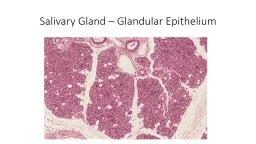
Examples: pancreatic acinar cells, serous cells in salivary glands
Slide72Serous secretory cells
Slide73Mucous secretory cellsAbundant rough ER, Golgi and secretory granules
Produce mucins (protective, lubricant glycoproteins)
Examples: mucous cells in stomach, goblet cells in small and large intestine, and mucous cells in salivary glands
Slide74Goblet cells in small intestine
Slide75Mucous secretory cells
Slide76Neuroendocrine cells
Dense secretory granules in cytoplasm contain polypeptides and/or amines (like epinephrine and norepinephrine)
Scattered throughout the body
Slide77Myoepithelial cellsSpindle-shaped cells found in glandular epithelial between basal lamina and basal cytoplasm
Embrace gland acini like an “octopus on a rock”
Contain actin: contract and squeeze out secretory product
Slide78Epithelium Lecture OutlineFunction and types of epitheliumStructure of epitheliumTypes of covering/lining epithelium
Types of glandular epithelium