
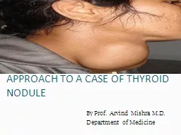
History The most common presentation for a thyroid nodule is A swelling noticed by the patient or by family and friends But if its not apparent how will it present Pressure symptoms is 1 way like dysphagia dyspnea stridor engorged neck veins or even ear pain and change in voice ID: 935985
Download Presentation The PPT/PDF document "Solitary thyroid nodule approach" is the property of its rightful owner. Permission is granted to download and print the materials on this web site for personal, non-commercial use only, and to display it on your personal computer provided you do not modify the materials and that you retain all copyright notices contained in the materials. By downloading content from our website, you accept the terms of this agreement.


Slide1
Solitary thyroid nodule
approach
Slide2History
The most common presentation for a thyroid nodule is?
A swelling noticed by the patient or by family and friends
But if its not apparent how will it present?
Pressure symptoms is 1 way like (dysphagia, dyspnea, stridor, engorged neck veins or even ear pain and change in voice)
Symptoms of hyper or hypothyroidism
(change in weight, heat or cold intolerance, change in bowel habits, sweating,…….so on)
Take history like normal history of onset, change in size, associated symptoms, pain
Slide3Our history should focus on
Any family history of thyroid disease
Drug history
And very important to ask about any history of radiation ( keeping in mind risk of papillary carcinoma is highly increased with radiation)
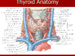
Slide4Now we exam our patient
Never forget to make sure is it thyroid? Or a neck swelling?
give the patient a sip of water if it moves upward on swallowing its attached to the thyroid, by?
Pretracheal
fascia
After confirming its of thyroid origin, we do our full exam of a mass first inspect, then palpate (percuss also), then auscultate)
And never miss any extra thyroid signs, neurological exam (reflexes, tremor)
LYMPHNODES!!!
Slide5Extrathyroid
?
Graves disease:
Exophthalmos
Lid retraction
Inflamed eyes
Double vision
Pretibial myxedema (non pitting, reddening and thickening of skin)
Lid lag
Hypothyroid:
Puffy face
Myxedema (nonpitting)
Dry skin, coarse hair
Bradycardia
Loss of lateral 1/3 of eyebrow
Hoarseness, slurred speech
Hyperthyroidism: tachycardia, palpitations and might even cause CHF
Slide6Before we continue lets take a minute
Goiters can be classified in different ways:
Benign or malignant
Simple or toxic
Diffuse or nodular (multinodular or solitary)
Slide7Investigate
Thyroid function tests
Measure free t3, t4, TSH
In thyrotoxicosis? TSH totally suppressed
In hypothyroidism? Elevated
Some things to keep in mind is in pregnancy or estrogen administration increases the level of thyroid hormone (increase thyroid binding globulin) in blood so it makes it harder to diagnose
So we use the T3 radioactive uptake
Slide8investigate
TRH and TSH stimulation tests
to determine the site of failure of production of thyroid hormone
Calcitonin levels are of importance too especially in diagnosis of medullary carcinoma
Lets not forget men2 syndrome
Men2A (medullary carcinoma of the thyroid, pheochromocytoma, parathyroid hyperplasia or adenomas)
Men2B (
medullay
carcinoma, pheochromocytoma, and neuromas (mucosal and intestinal)
Slide9Back to investigations
Thyroid antibodies
1-anti thyrocyte peroxidase antibody and anti thyroglobulin antibody (
hashimoto
thyroiditis)
2-thyroid stimulating immunoglobulin (graves disease)
And Radioisotope scanning (I123):
To differentiate between hot and cold nodules
If we have a solitary hot nodule it’s a toxic adenoma
If its cold we have multiple options (malignancy, benign, cyst)
ultrasound and FNA
FNA is best for discrete nodules
Slide10investigate
And we cant not mention MRI, CT, PET scan
But they aren’t in the routine assessment of a thyroid swelling
Mostly for assessment of a known malignancy, extent of a retrosternal mass, staging, or vascular invasion (MRI)
Now lets put things in a better way (more focused on a solitary nodule)
Slide11First keep in mind
Is benign or malignant?
Benign like: cyst, follicular adenoma (either toxic or simple),
thyroditis
Malignant like: medullary, follicular, papillary, anaplastic, maybe lymphoma)
Slide12So like we said history
So you’ve asked about everything we already said, family history,
radiation
, symptoms of hyper or hypo thyroid,…
Now u should pay attention to some stuff that might suggest a malignancy
Rapidly progressive
Young less than 15, or old over 65
Pain doesn’t suggest malignancy but if present doesn’t exclude malignancy (medullary cancer can cause dull aching pain)
Hoarseness is worrisome because it indicates malignant involvement of recurrent laryngeal nerve
If patient comes with painful thyroid you suspect subacute thyroiditis, so we ask about?
History of upper respiratory infection (virus) and fever
Pain in thyroid:
Medullary cancer
Bleeding in thyroid
Cyst
Subacute
thyroditis
Slide13Physical exam
On inspection or palpation we also have signs the should suggest to us malignancy
Firm
Fixed
Irregular margins
Cervical lymphadenopathy
Slide14investigate
Like we said our first investigation Is TFT, and this will direct us to what to do next
If the patient has low TSH it indicates that the nodule is secreting thyroid hormones on its own so we should further investigate by radio isotype:
If we get a hot nodule it’s a toxic adenoma (almost never malignant)
If its cold we should do ultrasound and FNA
On the other hand if we have normal or elevated TSH we don’t do radio isotype we go directly to ultrasound and FNA
Slide15Last thing:
ultrasound
Ultrasound often reveals multinodular goiters rather than solitary nodules, so to know the size and number of nodules
To know is it cystic or solid
And make a guess on how malignant is it
It can be used as guidance for FNA for accurate sampling
Might reveal features suggesting malignancy:
Microcalcifications
Irregular margins
Intra nodular vascular spots
Hypo echogenicity within the nodule
Slide16Management
Mainly depends on the cytology results from FNA
Malignancy needs surgical intervention depending on type of cancer
Benign lesions might be left alone and monitored if asymptomatic or surgically removed if symptomatic
About 30% of FNA turn out to be cysts and we just drain them, but re accumulation is common
We surgically remove cysts if its growing or painful
Slide17Slide18Slide19Slide20Slide21Slide22