

Alison Wright and Yaning Wang EN52048301SP20 BioPhotonics Laboratory Skin is the largest organ in the human body and consists of two principal layers the epidermis and the dermis The epidermis is a stratified squamous epithelium which consists of 4 types of cells3 ID: 931566
Download Presentation The PPT/PDF document "Multispectral Image Analysis for Skin Co..." is the property of its rightful owner. Permission is granted to download and print the materials on this web site for personal, non-commercial use only, and to display it on your personal computer provided you do not modify the materials and that you retain all copyright notices contained in the materials. By downloading content from our website, you accept the terms of this agreement.


Slide1
Multispectral Image Analysis for Skin Condition Diagnosis
Alison Wright and Yaning Wang
EN.520.483.01.SP20 Bio-Photonics Laboratory
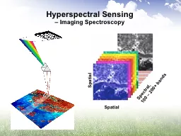
Slide2Skin is the largest organ in the human body and consists of two principal layers: the epidermis and the dermis. The epidermis is a stratified squamous epithelium, which consists of 4 types of cells:[3]Melanocytes, Langerhans cells, Merkel cells and Keratinocytes
Malignant melanoma is one of the most rapidly increasing cancers all over the world, with the estimated new cases of 76, 380 and estimated death of 10, 130 in the United States in 2016[1]
Clinical Background and Need
Fig.1&2 Anatomy of the skin and images of in
sinu melanoma(a), invasive melanoma(b)[3]
Slide3Dermoscopy is a popular in vivo non-invasive imaging tool that uses polarized light to aid dermatologists in examining pigmented skin lesions based on a set of morphological features.
Automatically segmenting melanoma from the surrounding skin is an essential step in computerized analysis of dermoscopic images.
Clinical Background and Need
Fig.3&4Challenges of automated lesion segmentation[1][2]
Fig.5
Dermoscopy
application
Slide4Unsupervised color image segmentation[4]Principal Component Analysis (PCA) Thresholding-based methods rely on the histogram distribution of image color which may be altered by hair and bubble in
RoI[1]..Classic GVF : influenced by distractions or noiseAbove methods all employ hand-crafted features that require specialized domain
knowledge
Previous work: automatic skin segmentation
Table.1 Comparison of several unsupervised segmentation methods
Slide5Supervised image segmentation method using convolutional neural networks These models have the capability of learning hierarchical features from raw image data without hand-crafted features.
Requires thousand annotated training samples
Previous work: automatic skin segmentation
Fig.6 Structure of unet
medical image segmentation[6]
Fig.7 Image mapping from cell membrane to segmentation map[6]
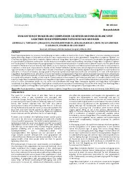
Slide6The use of skin color in dermatological diagnosis relies mainly on RGB (red, green, blue) color photography. This approach has limits due to the poor color discrimination of the human eye and brain. Multispectral imaging systems have the capacity to acquire images at different spectral bands, including wavelengths which the human eye is unable to capture.
Previous work: Multispectral imaging
Fig.8&9 Different multispectral imaging system[5][7]
Slide7Image preprocessing : hair removal and skin pore removalImage fusion for multispectral skin images based on wavelet decompositionSkin Lesion Segmentation using Bi-Directional
ConvLSTM U-Net Method
Slide8Hair removal – Edge detection & region filling Modify image preprocessing method[9]Example from ISIC 2016 dataset
Image preprocessing
Slide9Image registration(rigid transform) Image fusion(wavelet based)[8]Dataset:Oliver Lezoray University of Caen, IUT Grand Ouest Normandie, MMI Dpt.
Dermoscopy Skin Lesion Multispectral Image Database30 multispectral dermoscopic images (800 x 600) composed of 6 spectral bands (3 in visible light and 3 in infrared -IR- light)
Image fusion for multispectral system
Fig. 10&11 Structure of image fusion algorithm[7][8]
Slide10Image fusion result
Image fusion for multispectral system
near infrared reference visible light
near infrared reference visible light
Slide11Bi-Directional ConvLSTM U-Net[10]
Network architecture
Replace
Replace
Slide12Bi-Directional ConvLSTM U-Net[10]Bi-Directional
ConvLSTM U-NetLoss function: binary_crossentropySoftware: Keras,
Tensorflow-GPU backened 30 epoch batch_size 16Runtime: 40 minutes
Input data/Groundtruth: ISIC 2016 Challenge Part 1 500/200/200 training/validation/testing After image preprocessing
Network architecture
Slide13Result : input/
groundtruth/segmented result
Result
Slide14Evaluation compared with participants in ISIC 2016 challenge
Result
Participant
Accuracy
Jaccard Index
Sensitivity
Specificity
Urko
Sanchez
0.950
0.832
0.901
0.960
Mahmudur Rahman
0.949
0.884
0.859
0.962
Our
0.932
0.932
0.723
0.991
Slide15[1] Y. Yuan, M. Chao and Y. Lo, "Automatic Skin Lesion Segmentation Using Deep Fully Convolutional Networks With Jaccard Distance," in IEEE Transactions on Medical Imaging, vol. 36, no. 9, pp. 1876-1886, Sept. 2017.[2] A. Wong, J.
Scharcanski and P. Fieguth, "Automatic Skin Lesion Segmentation via Iterative Stochastic Region Merging," in IEEE Transactions on Information Technology in Biomedicine, vol. 15, no. 6, pp. 929-936, Nov. 2011.[3] K. Korotkov
and R. Garcia, “Computerized analysis of pigmented skin lesions: A review,” Artif. Intell. Med., vol. 56, no. 2, pp. 69–90, 2012[4]
Hance G, Umbaugh S, Moss R, Stoecker W. Unsupervised color image segmentation: with application to skin tumor borders. IEEE Engineering in Medicine and Biology 1996;15(1):104–11.[5] Romuald
Jolivot, Pierre Vabres, Franck Marzani,Reconstruction of hyperspectral cutaneous data from an artificial neural network-based multispectral imaging system,Computerized
Medical Imaging and Graphics,Volume 35, Issue 2,2011,Pages 85-88,ISSN 0895-6111,[6] Olaf RonnebergerEmail authorPhilipp FischerThomas
Brox, U-Net: Convolutional Networks for Biomedical Image Segmentation,MICCAI 2015 pp 234-241[7] V. Lukinsone, I. Kuzmina, R. Veilande, E. V. Plorina, D. Bliznuks, K. Bolochko, A. Derjabo, I. Lihacova, J. Spigulis, "Multispectral and autofluorescence RGB imaging for skin cancer diagnostics," Proc. SPIE 11065, Saratov Fall Meeting 2018: Optical and Nano-Technologies for Biology and Medicine, 110650A [8] Dong Han, Zhenhua Guo and D. Zhang, "Multispectral palmprint recognition using wavelet-based image fusion," 2008 9th International Conference on Signal Processing, Beijing, 2008, pp. 2074-2077[9] Okuboyejo D, Olugbara O, Odunaike S. Unsupervised restoration of hairoccluded lesion in dermoscopic images. In: Medical Image Understanding and Analysis Conference (MIUA ‘2014); London, UK. 2014. pp. 91-96 [10] R. Azad, M. Asadi, Mahmood Fathy and Sergio Escalera "Bi-Directional
ConvLSTM
U-Net with Densely Connected Convolutions ", ICCV, 2019
Reference
Slide16Thank you!