
دايمان سعود خليفة Types patterns of inflammatory response to infection 1 Suppurative inflammation 2 Granulomatous inflammation 3 Cytopathic cytoproliferative ID: 934028
Download Presentation The PPT/PDF document "Pathology of infectious diseases lab" is the property of its rightful owner. Permission is granted to download and print the materials on this web site for personal, non-commercial use only, and to display it on your personal computer provided you do not modify the materials and that you retain all copyright notices contained in the materials. By downloading content from our website, you accept the terms of this agreement.


Slide1
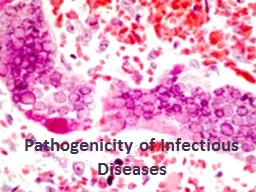
Pathology of infectious diseases lab
د.ايمان سعود خليفة
Slide2Types ( patterns ) of inflammatory response to infection:
1-
Suppurative
inflammation.
2-
Granulomatous
inflammation.
3-
Cytopathic
/
cytoproliferative
.
4- Necrotizing inflammation.
5- Chronic inflammation.
Slide3Suppurative infl. Lung
Slide4Granulomatous inflammation.
Slide5Slide6Epitheloid cells
Slide7Slide8Cytopathic-cytoproliferative response
Slide9inclusion bodies
in Cytomegalovirus
Slide10A stained cell infected with cytomegalovirus. Note the enlarged nucleus (purple) and the virus inside inclusion bodies (darker pink)
Slide11Slide12Necrotizing soft tissue infection
Slide13Bacterial infectionStaphylococcal
Streptococcal
cholera
T.B
Leprosy
Syphilis
Slide14Slide15Slide16Slide17Slide18Streptococcal infections:1)
Suppurative
diseases:
Pneumonia, cellulites , erysipelas
2)
Nonsuppurative
diseases
Slide19cellulitis
Slide20erysipelas
Slide21Scarlet fever
Slide22Group A βeta –hemolytic
Streptococcal infection
Responsible for post-infectious syndromes:
rheumatic fever
poststreptococcal
glomerulonephritis
Slide23T.B:1)Primary T.B 2) Progressive primary T.B
3)Secondary T.B
Slide24Slide25Slide26Slide27Slide28Ziehl–Neelsen stain
Slide29Secondary T.B
Primary T.B
Adult
Children
Age
Exogenous or endogenous (reinfection from the primary site )
Exogenous exposure to m.o
Source
Apex of the lung + extrapulmonary (intestine, bone kidney , adrenal)
Subpleural region of the lung
Site
The pulmonary lesion is larger and more cavitating than the L.N lesion.
The L.N lesion is larger than the pulmonary lesion.
Lymph node
Positive
Negative
Mantoux test
Symptomatic (fever, night sweat, hemoptysis.
The patient is infectious to others and excrete the bacilli into the environment.
Majority are Asymptomatic, 90% heal.
Course of the dis.
Slide30Interpretation of tuberculin (mantoux) test:
Slide31Slide32Slide33Leprosy1) Lepromatous2)
Tuberculoid
3) Borderline
Slide34Leonine face
Slide35Slide36Slide37Syphilis1)congenital2)aquired
Primary
Secondary
tertiary
Slide38Slide39Saddle nose
Slide40Saber shine deformity
Slide41Syphilitic chancre
Slide42Slide43Secondary syphilis macular rash
Slide44Slide45Condylomata lata
condyloma
accuminata
Slide46Tertiary syphilis Gumma of the face
Slide47Slide48Gumma of the liver
hepar
lobatum
Slide49Slide50DX of syphilis:
Direct examination:
dark field
illumination &
immunoflourescence
microscopy
+
ve
in primary and secondary syphilis
Serology:
NOT specific to syphilis:
VDRL
Wasserman's reaction.
specific for syphilis:
TPIT
TPHAT
+
ve
in tertiary syphilis
Slide51Slide52CUTANEOUS LEISHMANIA
Slide53MUCOCUTANEOUS LEISHMANIA
Slide54Leishmania donovani
Slide55Rupture of the macrophage
Slide56Entamoeba histolytica
Colonic lesion
Hepatic lesion
Slide57Slide58Mic.of flask shape ulcer
Slide59Hydatid cyst
Slide60Slide61Slide62Slide63Swimmer’s itch
Slide64Slide65Slide66Slide67aspergilloma
Slide68Slide69Slide70Thank you