
Akash Srinivasan as14317icacuk Shortness of Breath Heart Failure Cardiomyopathy Constrictive pericarditis Myocarditis Shortness of Breath Poor removal of CO2 Poor delivery of oxygen ID: 932829
Download Presentation The PPT/PDF document "Cardiac Causes of Shortness of Breath" is the property of its rightful owner. Permission is granted to download and print the materials on this web site for personal, non-commercial use only, and to display it on your personal computer provided you do not modify the materials and that you retain all copyright notices contained in the materials. By downloading content from our website, you accept the terms of this agreement.


Slide1
Cardiac Causes of Shortness of Breath
Akash Srinivasanas14317@ic.ac.uk
Slide2Shortness of Breath
Heart Failure
Cardiomyopathy
Constrictive pericarditis
Myocarditis
Slide3Shortness of Breath
Poor removal of CO2
Poor delivery of oxygen
Slide4Shortness of Breath
1. Not enough oxygen reaching the lungs
Breathing issues
E.g. asthma, COPD, anaphylaxis
Slide5Shortness of Breath
2. Not enough oxygen getting into the blood
V/Q mismatch
E.g. pulmonary embolism, pulmonary oedema, pulmonary fibrosis
Slide6Shortness of Breath
3. Not enough oxygen reaching the rest of the body
Heart issues
(or anaemia, shock etc.)
Slide7Shortness of Breath
Heart Failure
Cardiomyopathy
Constrictive pericarditis
Myocarditis
Slide8SBA 1
JB is a 34-year-old male, with a history of infective endocarditis, complaining that he’s tired all the time and struggles to run as far as he used to. He also says that his ankles and face feel more swollen than before. On examination, he has a raised JVP, breathing rate and heart rate. You also hear a pansystolic murmur on auscultation. What is the most likely diagnosis?
A. Left heart failure secondary to mitral regurgitation B. Left heart failure secondary to cocaine abuse C. Right heart failure secondary to tricuspid regurgitation
D. Myocardial infarction
E. High output heart failure
Menti: 15 04 90 5
Slide9Definition:
The failure of the heart to maintain the cardiac output (CO) required to meet the body’s
demands
Not enough oxygen reaches the rest of the body
Heart Failure
Slide10HF: Classification
Slide11HF: Classification
Left Heart Failure
(LHF)
Right Heart Failure
(RHF)
LHF + RHF =
Congestive Heart Failure
(CHF)
Slide12HF: Classification
Low Output State: Heart fails to pump in response to normal exertion -> low CO
High Output State: CO is normal but higher metabolic needs e.g. pregnancy, anaemia, hyperthyroidism
Slide13Chronic Left HF: Aetiology
ValvularAortic stenosisAortic regurgitation
Mitral regurgitationMuscular
Ischaemia (IHD)
Cardiomyopathy
Myocarditis
Arrhythmias (AF)
SystemicHypertensionAmyloidosisDrugs (e.g. cocaine, chemo)
Slide14Chronic Right HF: Aetiology
LungsPulmonary hypertension (cor pulmonale)
Pulmonary embolismChronic lung disease e.g. interstitial lung disease, cystic fibrosis
Valvular
Tricuspid regurgitation
Pulmonary valve disease
LHF -> CHF
Slide15Chronic High Output HF: Aetiology
Conditions that require a higher CO = strain on the heart NAP MEALSNutritional (B1/thiamine deficiency)
Anaemia
P
regnancy
M
alignancyEndocrine
AV malformationsLiver cirrhosisSepsis
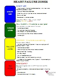
Slide16HF: Signs and Symptoms
What happens if fluid is congested backwards?
Slide17LHF: Symptoms
Respiratory ProblemsDyspnoea: Paroxysmal nocturnal dyspnoea (PND)
Exertional dyspnoea
Orthopnoea
Nocturnal cough
(+/- pink frothy sputum)
Fatigue
OSCE TIPS:1) Assess SOB: “How far are you able to walk before getting breathless? How many flights of stairs?”2) Assess orthopnoea: “Have you noticed anything making the SOB worse? What about lying down, standing up?” 3) Assess PND: ”Do you ever wake up at night gasping for air? How many pillows do you sleep with at night? Has this changed recently?”
Slide18LHF: Signs
Heart
Lungs
↑HR, ↑RR
Irregularly irregular heartbeat
Pulsus alternans
Displaced apex beat
S3 Gallop rhythmS4 in severe HFMurmur (AS, MR, AR)Fine end-inspiratory crackles at lung bases (pulmonary oedema)Wheeze (cardiac asthma)
Slide19RHF: Signs and Symptoms
SymptomsFatigueReduced exercise tolerance
AnorexiaNausea
Nocturia
Signs
Face
: face swelling
Neck: ↑ JVPHeart/Chest: TR murmur, ↑ HR, ↑ RRAbdomen: ascites, hepatomegalyOther: ankle and sacral pitting oedema
Slide20HF: Summary of Signs and Symptoms
Slide21SBA 1
JB is a 34-year-old male, with a history of infective endocarditis, complaining that he’s tired all the time and struggles to run as far as he used to. He also says that his
ankles and face feel more swollen
than before.
On examination, he has a
raised JVP
, breathing rate and heart rate. You also hear a pansystolic murmur on auscultation. What is the most likely diagnosis?
A. Left heart failure secondary to mitral regurgitation B. Left heart failure secondary to cocaine abuse C. Right heart failure secondary to tricuspid regurgitation D. Myocardial infarction E. High output heart failure
Slide22SBA 1
A. Left heart failure secondary to mitral regurgitation No breathlessness symptoms – LHF unlikely B. Left heart failure secondary to cocaine abuse No breathlessness symptoms – LHF unlikely
C. Right heart failure secondary to tricuspid regurgitation
Signs of RHF, pansystolic murmur, infective endocarditis
D. Myocardial infarction This would present more acutely with crushing chest pain
E. High output heart failure No mentioned condition like anaemia or hyperthyroidism
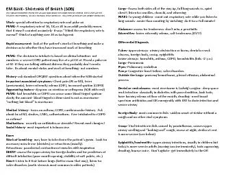
Slide23HF: Investigations
Slide24HF: Investigations
↓
BNP
↑BNP
HF unlikely
TTE
BNP is SENSITIVE but
NOT SPECIFIC
Slide25HF: Investigations
Transthoracic echocardiogram (TTE) coupled with doppler = DIAGNOSTICVisualise the structure and function of the heart -> may show the cause of HF
Can calculate ejection fraction (EF): % of the blood present in the LV that gets pumped during systole – normal = 50-70%
EF < 40%:
HF with reduced ejection fraction (
HFrEF
) – previously called
systolic HFIndicates inability of the ventricle to contract normallyEF >50%: HF with preserved ejection fraction (HFpEF) – previously called diastolic HFIndicates inability of the ventricle to relax and fill normally
Slide26HF: Investigations
Chest X-Ray Alveolar oedema
B-lines (
Kerley
)
C
ardiomegaly
Dilated upper lobe vessels + Diverted upper lobe Effusion (Transudative pleural effusion)
Slide27HF: Diagnosis
Clinical diagnosis can be made using the Framingham Criteria 2+ majors OR 1 major and 2 minors
Major:
Paroxysmal nocturnal dyspnoea
Bibasal crepitations
S3 gallop
Cardiomegaly
Increased central venous PressureWeight lossNeck vein distensionAcute pulmonary oedemaHepatojugular reflux Minor- bilateral ankle oedema- dyspnoea on ordinary exertion- tachycardia
- decrease in vital capacity by 1/3
- nocturnal cough
- hepatomegaly
- pleural effusion
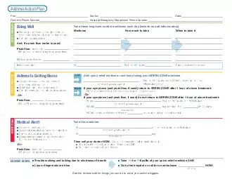
Slide28HF: Management
For Chronic HF:
Slide29HF: Management
ACE inhibitors: give to all patients with LV dysfunctionEnalapril, perindopril, ramipril (all end in –pril)Can switch to ARB if not tolerable (cough)
Beta-blockers: reduce O2 demand on the heartBisoprolol, carvedilol
Diuretics: use if evidence of fluid retention
Loop diuretics e.g. furosemide
Aldosterone antagonists e.g. spironolactone
Hydralazine + nitrates – considered in Afro-Caribbean patients Digoxin – positive inotrope, improves symptoms
but not mortality Cardiac resynchronization therapy – aims to improve timings of contraction of atria and ventricles
Slide30HF: Management
DMONSDiureticsMorphine
OxygenNitratesS
it-up
For Acute HF:
MEDICAL EMERGENCY - ABC
Slide31HF: Complications and Prognosis
Complications:Respiratory failureRenal failure - due to hypoperfusion
Acute exacerbations
Death
Prognosis:
Very poor, worse than most malignancies
50% of severe HF patients die within 2 years
Acute HF in-hospital mortality = 2-20%
Slide32SBA 2
After measuring BNP levels and performing echocardiography on JB, a diagnosis of heart failure is confirmed. You perform a chest X-ray as well. What would you expect to see on the CXR? A. Reduced cardio-thoracic ratio B. Lower lobe diversion
C. Sharp costophrenic angles D. Kerley C lines E. Alveolar oedema
Menti: 15 04 90 5
Slide33SBA 2
After measuring BNP levels and performing echocardiography on JB, a diagnosis of heart failure is confirmed. You perform a chest X-ray as well. What would you expect to see on the CXR?
A. Reduced cardio-thoracic ratio B. Lower lobe diversion C. Sharp costophrenic angles D. Kerley C lines
E. Alveolar oedema
Slide34SBA 2
A. Reduced cardio-thoracic ratio Cardiomegaly so cardio-thoracic ratio is increased B. Lower lobe diversion
Upper lobe diversion
C. Sharp costophrenic angles
Pleural effusion results in blunt costophrenic angles
D. Kerley C lines
Kerley B lines E. Alveolar oedema Left heart failure causes pulmonary oedema
Slide35Shortness of Breath
Heart Failure
Cardiomyopathy
Constrictive pericarditis
Myocarditis
Slide36Definition:
A group of diseases in which the myocardium
becomes structurally and functionally abnormal
In the absence of coronary artery disease, valvular disease and congenital heart disease
It can affect young people
Cardiomyopathy
Slide37Cardiomyopathy: Types
Slide38Cardiomyopathy: General Presentation
HistorySymptoms of HF:SOB on exertion
FaintingFatigue
Sudden death often 1
st
presentation
Family history
ExaminationSigns of HF:Respiratory cracklesMurmursS3, S4
Investigations
No single diagnostic test for all types
ECHO
Can also do bloods, BNP, CXR, ECG, cardiac catheterisation, stress test
OSCE TIP:
Ask if there is a family history of sudden, unexplained cardiac death at a young age e.g. under 50.
Slide39Dilated Cardiomyopathy
Pathophysiology Ventricles enlarge and become dilated. Walls thin and weaken -> can’t contract effectively. Think of the Law of Laplace: increased radius leads to reduced ventricular pressure
Risk Factors
Alcohol
, post-viral, haemochromatosis, genetic
Presentation
Signs and symptoms of HF
Displaced apex beat
, TR/MR murmur, S3
Investigations
Globular heart on CXR, dilated ventricle on Echo
Slide40Hypertrophic Cardiomyopathy
PathophysiologyH for Hench – muscle thickens inwards.
Increased stiffness of the muscle affects pumping.Thickened muscle disrupts electrical conduction and causes arrythmia.
Hypertrophic Obstructive Cardiomyopathy (HOCM) = thickened ventricle obstructs the outflow of blood.
50% is familial (autosomal dominant)
Slide41Hypertrophic Cardiomyopathy: Presentation
Symptoms
Usually asymptomatic
Sudden cardiac death is often the 1
st presentation Angina, dyspnoea on exertion, palpitations, syncope Signs Ejection systolic murmur Jerky carotid pulse Double apex beat but NOT DISPLACED S4
Investigations
ECG: Q waves, left axis deviation, signs of left ventricular hypertrophy
Echo: ventricular hypertrophy (asymmetrical septal hypertrophy
Amir Sam’s Tip:
LVH by voltage criteria:
Deep S in V1/2
Tall R in V5/6
S in V1 + R in V5 or V6
≥ 7 large squares
Slide42Restrictive Cardiomyopathy
PathophysiologyR for Rigid – ventricles become abnormally rigid and lose flexibility.Impaired ventricular filling during diastole.
Reduced preload -> reduced blood flow + backing up of blood.
Causes
Sarcoidosis, amyloidosis, haemochromatosis (the infiltrative ”osis” diseases)
Familial
IdiopathicRarer than dilated or hypertrophic cardiomyopathy
Symptoms
Asymptomatic or HF symptoms
Signs
RHF signs: raised JVP, S3, ascites and oedema, hepatomegaly
Kussmaul’s sign = paradoxical rise in JVP during inspiration
Slide43Other Cardiomyopathies
Arrhythmogenic Right Ventricular CardiomyopathyProgressive fatty and fibrous replacement of the ventricular myocardiumInherited (autosomal dominant)
Takotsubo Cardiomyopathy
Sudden temporary weakening of heart muscle after a
significant stressor
”Broken heart syndrome”
Slide44Shortness of Breath
Heart Failure
Cardiomyopathy
Constrictive pericarditis
Myocarditis
Slide45Constrictive Pericarditis
Definition
Chronic inflammation of the pericardium (outer sac) with thickening and scarring
Causes
Idiopathic
Infectious (TB, bacterial, viral)
Acute pericarditis
Cardiac surgery and radiation
Signs and Symptoms
Similar to restrictive cardiomyopathy
RHF presentation (raised JVP, oedema)
Kussmaul’s sign
Investigations
CXR: pericardial calcification
Echo: increased pericardial thickness – differentiate from restrictive cardiomyopathy
Cardiac CT/MRI
Slide46Shortness of Breath
Heart Failure
Cardiomyopathy
Constrictive pericarditis
Myocarditis
Slide47Myocarditis
Definition
Inflammation of the myocardium
Inflammatory cardiomyopathy
Causes
Infectious
Drugs - cocaine
Metals
Radiation
Coxsackie B virus is the most common cause of myocarditis in Europe
Signs and Symptoms
Flu-like prodrome
Chest pain (worse when lying down)
SOB
Palpitations
Investigations
ECG: non-specific ST and T wave changes
Cardiac biomarkers: CK and troponin
Endomyocardial biopsy:
diagnostic but not routinely performed
Slide48Shortness of Breath
Heart Failure
Cardiomyopathy
Constrictive pericarditis
Myocarditis
Slide49SBA 3
Aston was a 33-year-old male who suddenly collapsed on stage. Although the doctors attempted “love CPR”, the patient died, and the post-mortem revealed a hypertrophic heart. What was the most likely cause of death? A. Obstructed flow of blood from the heart B. Arrhythmia
C. Reduced pumping of blood due to stiff myocardium D. Stroke E. Sub-arachnoid haemorrhage
Menti: 15 04 90 5
Slide50SBA 3
Aston was a 33-year-old male who suddenly collapsed on stage. Although the doctors attempted “love CPR”, the patient died, and the post-mortem revealed a
hypertrophic heart. What was the most likely cause of death?
A. Obstructed flow of blood from the heart
B. Arrhythmia C. Reduced pumping of blood due to stiff myocardium D. Stroke E. Sub-arachnoid haemorrhage
Slide51SBA 3
A. Obstructed flow of blood from the heart Likely to experience warning symptoms beforehand B. Arrhythmia
Most likely cause of death from HCM, hypertrophic muscle affects electrical circuits
C. Reduced pumping of blood due to stiff myocardium
Likely to experience warning symptoms beforehand
D. Stroke Heart issue rather than brain issue, ventricular arrhythmia (not AF)
E. Sub-arachnoid haemorrhage Heart issue rather than brain issue
Slide52SBA 4
Oritsé presents with breathlessness and he says that he experienced a fever recently. His CK and troponin are elevated, so a presumptive diagnosis of myocarditis is made. What other signs or symptoms would be consistent with this diagnosis?
A. Kussmaul’s sign B. Ankle oedema C. Ascites D. Jaundice E. Chest pain
Menti: 15 04 90 5
Slide53SBA 4
Oritsé presents with breathlessness and he says that he experienced a fever recently. His
CK and troponin are elevated, so a presumptive diagnosis of myocarditis is made.
What other signs or symptoms would
be most
consistent with this diagnosis?
A. Kussmaul’s sign B. Ankle oedema C. Ascites D. Jaundice
E. Chest pain
Slide54SBA 4
A. Kussmaul’s sign B. Ankle oedema C. Ascites D. Jaundice
All of these are subacute/chronic signs of heart failure, restrictive cardiomyopathy, constrictive pericarditis etc. E. Chest pain
Only option which fits with the acute picture of myocarditis
Slide55Scan here
Before the final question…
Slide56SBA 5
Marvin presents with a 4-month history of increasing breathlessness and ankle swelling. On examination, he has ascites and Kussmaul’s sign is elicited. What would be the most useful diagnostic investigation? A. Echocardiography B. ECG
C. Endomyocardial biopsy D. Abdominal X-ray E. CK
Menti: 15 04 90 5
Slide57SBA 5
Marvin presents with a 4-month history of increasing breathlessness and ankle swelling. On examination, he has ascites
and Kussmaul’s sign is elicited. What would be the
most useful diagnostic investigation
?
A. Echocardiography B. ECG C. Endomyocardial biopsy D. Abdominal X-ray E. CK
Slide58SBA 5
A. Echocardiography Allows differentiation between restrictive cardiomyopathy and constrictive pericarditis
B. ECG Non-specific signs – not the most useful
C. Endomyocardial biopsy
Pericardial biopsy might be useful – but highly invasive
D. Abdominal X-ray Chest X-ray would be useful to look for pericardial calcifications, but these are not specific to constrictive pericarditis E. CK May be mildly elevated in both constrictive pericarditis and restrictive cardiomyopathy – not that helpful
Slide59Any Questions?
Email me any questions: as14317@ic.ac.uk Please fill in the feedback
:)
Scan here