
J Ochotná The main functions of the immune system Immune system belongs to the basic homeostatic mechanisms Defense Autotolerance Immune ID: 931203
Download Presentation The PPT/PDF document "http://uia.fnplzen.cz/ Immune system" is the property of its rightful owner. Permission is granted to download and print the materials on this web site for personal, non-commercial use only, and to display it on your personal computer provided you do not modify the materials and that you retain all copyright notices contained in the materials. By downloading content from our website, you accept the terms of this agreement.


Slide1
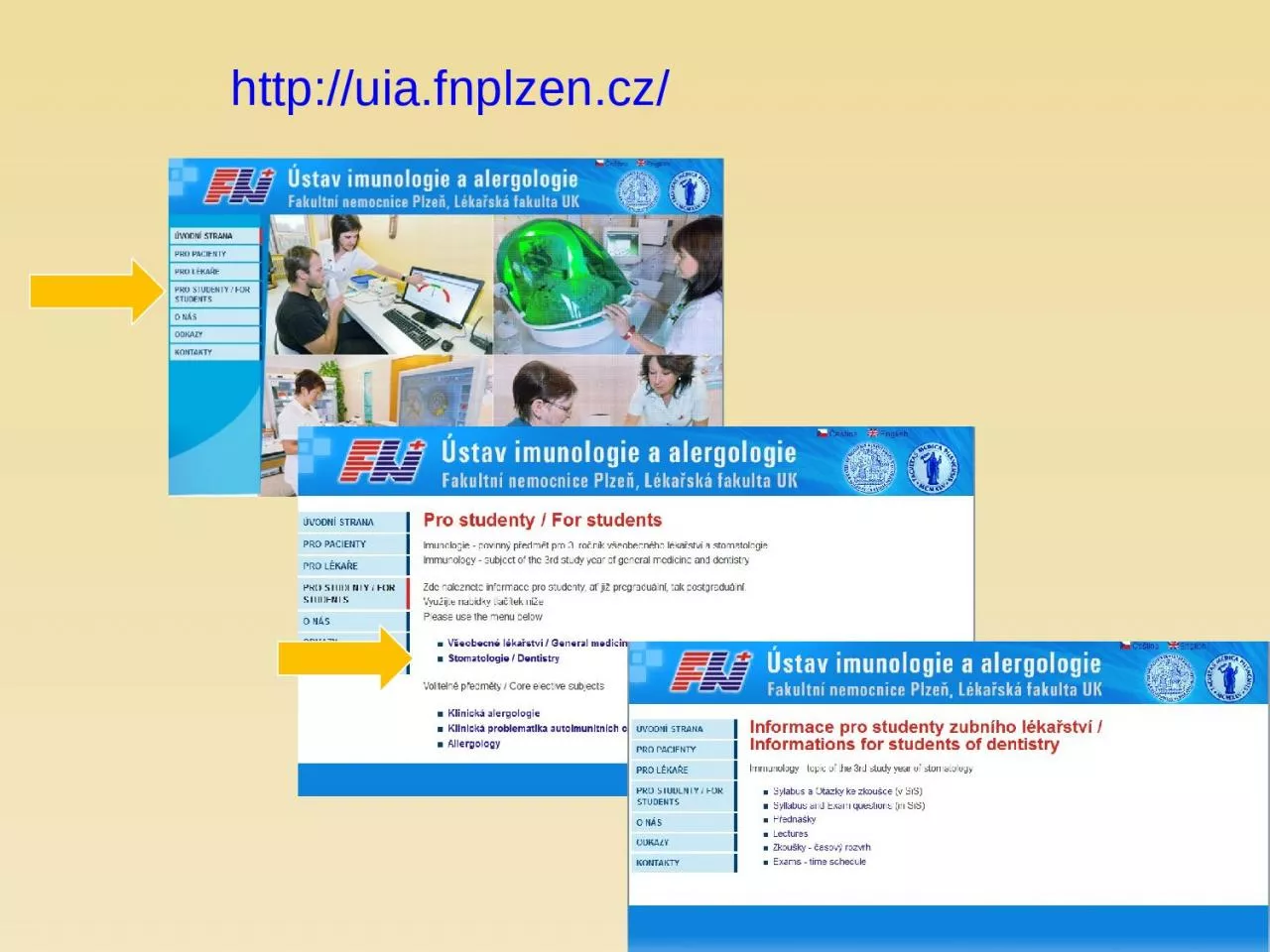
http://uia.fnplzen.cz/
Slide2Immune system
J. Ochotná
Slide3The
main functions
of
the immune system
Immune
system
belongs
to
the
basic
homeostatic
mechanisms
Defense
Autotolerance
Immune
surveillance
Slide4Antigen (
immunogen)
* substance
that can
induce a
humoral
and/or cell-mediated immune
response
*
predominantly
proteins
or
polysaccharides
*
molecules
>5
kDa
(
optimal
size
of
antigen
is
about
40
kDa
)
*
autoantigen
*
exoantigen
*
allergen
Slide5Haptens
*
small
molecules, that are able
to induce
specific
immune
response
only after the establishment to the macromolecular carrier (separate haptens are not immunogenic) * typically drugs (eg penicillin antibiotics, hydralazin)
Slide6Interaction
antigen – antibody
*
Binding
site of
antibody
(
paratop
) form non-covalent complexes with the corresponding part on antigen molecule (epitope)* antigen-antibody complex is reversible
Slide7Types
of antigens
according to antigen
presentation1)
thymus dependent
antigens
*
more
frequent, especially protein Ag* for induction of humoral immune response is necessary cooperation with TH
cells
(
or
response isn´t enough effective)* assistance implemented in the form of cytokines produced by TH cells
Slide8Types
of antigens
according to antigen
presentation2)
thymus independent antigens
*
can
induce antibodies production directly without the participation of T lymphocytes* mainly bacterial polysaccharides,
lipopolysaccharides
and
polymer forms of proteins(e.g. Haemophilus, Str.pneumoniae)T-independent pathway
Slide9Superantigens
* stimulate
lymphocytes
polyclonaly
and
massively
(5-20%)
* massive activation of T lymphocytes can cause shock* e.g. bacterial toxins (Staph.aureus, Str.pyogenes, Pseud.aeruginosa)
Slide10Sequestered
antigens
*
autoantigens that are normally
hidden
from
the
immune
system and therefore unknow (e.g. the lens of the eye , testes, brain) * if they are "
uncovered
" by
demage
, can induce the immune response (one of the theories of autoimmune processes)
Slide11Components
of the
immune
system
Slide12Components
of the
immune
system* Lymphoid
tissues and
organs
*
Cells
of the immune system* Molecules of the immune system
Slide13Lymphoid
tissues and
organs
* are linked with
the other
organs
and
tissues by network of lymphatic and blood vessels Primary lymphoid tissues and organs* bone marrow, thymus* maturation and differentiation of immunocompetent
cells
*
immature lymphocytes acquire here their antigenic specificity
Slide14Secondary
lymphoid tissues
and
organs * meeting
place of
immunocompetent
cells
with
Ag spleen - filters the blood and captures presented antigenslymph nodes and their organized clusters (tonsils, appendix, Peyer patches in the intestine) - filter
lymph
and capture present antigens MALT (mucous associated lymphoid tissue) - diffuse lymphatic tissue, the main role is capture of antigens passing through
the
mucosal
epithelium
Slide15Cells
of the
immune
system*
development
of
red
and
white blood cells begin at yolk sack, then haematopoiesis travels to fetal liver and spleen (3 to 7 month gestation),
then
bone
marrow has the main hematopoietic function * all blood cells arise from a pluripotent stem cell (CD 34)
*
haematopoiesis
is
regulated
by
cytokines
Slide16Slide17Immune
mechanisms
Slide18Nonspecific
(innate) immune
mechanisms
* non-adaptive,
innate
*
evolutionarily
older
* no immunological memory* in the presence of pathogens react quickly, in minutes (based on molecules and cells
which
are in
the
body prepared in advance)* component cellular – granulocytes (neutrophils, eosinophils, basophils), monocytes (macrophages, DC), NK cells
, mast
cells
humoral
-
complement
,
interferons
,
lectins
and
other
serum
proteins
Slide19Specific
(adaptive) immune
mechanisms
* adaptive
, antigen-specific
*
evolutionarily
younger
* have immunological memory* development of a full-specific immune response takes several days even
weeks
*
component cellular - T lymphocytes (TCR) humoral - antibodies
Slide20Phagocytosis
Slide21Phagocytosis
=
ability to absorb
particles from the
surroundings
Professional
phagocytes
*
cells, which provide defenses by mechanism of phagocytosis * neutrophilic granulocytes, monocytes, macrophages and DC
Slide22Professional
phagocytesgranulocytes - defense
against extracellular
pathogens - able to
perform effector
functions
immediately
macrophages
- removal of own apoptotic cells, defense against certain intracellular parasites, APC - fully functional after activation by cytokines (IFNg, TNF)
Macrophage
Slide23The
migration of
phagocytes
in damaged and
infected tissues
7%
of
peripheral
neutrophils and phagocytes93% neutrophils and phagocytes in the bone marrow* in place of damage phagocytes are captured
on
endothelium
(
due to inflammatory cytokine expression of adhesion molecules is higher)
Slide24Phagocytosis
*
the
first interactions with
adhesion
molecules
slows
the
movement of neutrophils - roling * then there is a stronger link between endothelial cells and leukocytes and subsequent
penetration
between
endothelial cells to the tissue - diapedesis (or extravasation)* phagocytes direct their movement to the
site
of
inflammation
by
chemokines
(IL-8, MIP-1
a
and
b
, MCP-1, RANTES,
C3a
, C5a,
bacterial
products
...)
Slide25Slide26Receptors
on phagocytes
PAMPs
(pathogen associated
molecular
patterns
)
PRR
(
pathogen
recognition receptors) * TLR receptors (binds bacterial lipoproteins, lipopolysaccharides, bacterial DNA) * mannose receptor* galactose receptor* CD14 (binds
bacterial
LPS
)* scavenger receptors (bind phospholipids on the surface of apoptotic cells)
Slide27Opsonisation
*
is the process by which a pathogen is marked for ingestion
and
d
estruction
by
phagocyte
* Opsonins - IgG, IgA, C3b, MBL, fibronectin, fibrinogen, CRP, SAP* Fc receptors on phagocytes (recognize antibodies linked
to
surface
of micro-organism)* complement receptors (for binding C3b)
Slide28Phagocytosis
Slide29Degradation
of ingested
material
*
fagosome
fusion
with
lysosomes
- oxygen independent(lysozyme, defensines, serine proteases, myeloperoxidase, acidic pH…) * activation of membrane NADPH oxidase - oxygen dependent (superoxide
,
hydrogen
peroxide,
hypochlorous acid)* production of nitric oxide (NO) by macophages
Slide30Secretory
products of
phagocytes
* IL-1, 6, TNF (systemic response to inflammation
)
* IL-8 (
chemokine
)
* IL-3, GM-CSF (
control
haematopoiesis)* TGFa, TGFb (tissue regeneration)* metabolic products of arachidonic acid (prostaglandins,
prostacyclin
,
leukotrienes and thromboxanes)
Slide31Complement
Slide32Complement
*
system of
about 30 serum and
membrane proteins
*
complement
components are present in the serum in inactive form* complement activation has cascade character* complement proteins are synthesized in the liver, less by tissue macrophages and
fibroblasts
*
the main complement components: C1-C9 (C3 is the central component)* other complement components: factor B, factor D, factor P*
regulatory
proteins
: C1 - inhibitor,
factor
I,
factor
H, DAF, MCP,
CR1, CD59 (
protektin
)
inactivator
of
anafylatoxin
…
Slide33Complement
functions
* Opsonization (C3b)
* Chemotaxis (C3a, C5a)
* Osmotic
lysis
(MAC C5b-C9)
*
Anafylatoxins
(C3a, C4a, C5a)
Slide34Complement
activation
*
Alternative pathway*
Clasial pathway
*
Lektin
pathway
Slide35Slide36Regulation
of complement
and
protection of
own
cells
Activation of complement cascade is controlled by the plasma and membrane inhibitors.
MCP
DAF
Protectin
Anaphylatoxin inactivator
Slide37Complement
regulation
C1 inhibitor
(C1-INH) – inhibits C1; if missing→ HAE
factor I
with cofactors:
MCP
(membrane cofactor protein),
CR1
,
factor H – C3b, C4b cleavageDAF (decay-accelerating protein)-degradation of C3 and C5 convertase
Slide38factor
S
(
vitronectin) – inhibits
complex C5bC6
CD 59
(
protectin
) -
prevents the polymerization of C9 anaphylatoxin inactivator (CPN)- inactivates anafylatoxins (C3a, C4a, C5a)Complement regulation
Slide39Complement
receptors
* Bind
fragments of
complement components
CR1 - on
various
cells
-
removing of immunecomplexesCR2 - on B lymphocytes and FDC - activation of B cellsCR3, CR4 - on phagocytes
-
participation
in opsonization, adhesion
Slide40Basophils
and mast cells
and
their importance in immune
responses
Slide41Mast
cells
Mucosal mast cells
- in the mucous membranes
of
respiratory
and
gastrointestinal
tract, participate in parasitosis and allergy Connective tissue mast cells - the connective tissue, in parasitosis and allergy are not participating
Slide42Mast cell
functions
Defense against
parasitic infections
Responsible
for
the
early type of hypersensitivity (allergic reaction)Apply during inflammation, in angiogenesis, in tissue remodeling Regulation of immune response
Slide43Mast cell
activation
Mast cells
degranulation can be
stimulated by:
cross
-
linking
of
IgE Fc receptorsanafylatoxins (C3a, C4a, C5a) TLR
Slide44Mast cell
activation by cross-linking
of
IgE Fc receptors
Establishing
of
multivalent antigen (
multicellular
parasite)
to IgE linked to highaffinnity Fc receptor for IgE (FcRI) Aggregation of several molecules FcRI Initiate mast cell degranulation (cytoplasmic granules mergers
with
the
surface membrane and release their contents) Activation of arachidonic acid metabolism (leukotriene C4, prostaglandin D2) Production of cytokines (TNF, TGF, IL-4, 5,6 ...)
Slide45Activation
schema of mast cell
Slide46Secretory
products of mast
cells
Cytoplasmatic granules:
hydrolytic enzymes
, heparin, chondroitin
sulphate
, histamine, serotonin
Arachidonic
acid metabolites (leukotriene C4, prostaglandin D2) Cytokines (TNF, TGF , IL-4, 5,6 ...)
Slide47Histamine
vasodilation
increased vascular permeability
bronchoconstrictionincreases
intestinal peristalsis
increased
mucus
secretion
Slide48Basophils
Differentiate
from myeloid
precursor
Are very similar
to mast
cells
by
the
receptor
equipment, content of granules, the mechanisms of stimulation and functionsPlay role in inflammation, regulation of immune responses, in allergic reactions, they are responsible for the emergence of anaphylactic shockIn high numbers at the sites
of
ectoparasite
infection
Slide49Slide50Complement
– clasical pathway
https://www.youtube.com/watch?v=vbWYz9XDtLwComplement –
alternative pathwayhttps://www.youtube.com
/watch?v=qga3Wn76d9w
Complement
https://www.youtube.com/watch?v=5Ao36HNvwvw
Immune
reaction
https://www.youtube.com/watch?v=G7rQuFZxVQQ
Slide51Alternative
complement pathway
* C3 component
of complement
spontaneously
breaks
into C3b and C3a* C3b can covalently bind on the surface of microorganism * to bound C3b join a factor B, which is cleaved by factor D to
Ba
and
Bb, resulting complex C3bBb is stabilized by factor P and functions as an alternative C3 convertase* C3 convertase cleaves C3 to C3a (chemotaxis) and C3b, which
binds
to
the
surface
of
the
microorganism
(
opsonization
),
or
gives
rise
to
other
C3 convertases*
from
some
C3
convertases
form
C3bBbC3b
that
act
as
an
alternative
C5
convertase
,
which
cleaves C5 to C5a (chemotaxis) and C5b (starts terminal lytic phase)
Slide52Classical
complement pathway
*
Can be
initiated by
antibodies
(
IgM
,
IgG
, except IgG4) or by CRP, SAP , which are bound to antigen* C1 binds to antibodies that have already attached themselves to antigen , change its conformation and get proteolytic activity - starts cleave
proteins
C4
and C2* fragments C4b and C2a bind to the surface of the cell and create the classic C3 convertase (C4bC2a), which cleaves C3 to C3a
and
C3b
*
then
creates
a
classic
C5
convertase
(C4bC2aC3b)
that
cleaves
C5
to
C5a
and
C5b
Slide53Lectin
complement pathway
*
is initiated
by serum
mannose
binding
lectin
(MBL)* MBL binds to manose, glucose or other sugars on the surface of some microbes, after the bindins starts cleave
C4
and
C2* this way is similar to the classical pathway
Slide54Terminal (
lytic) phase of
the
complement cascade
C5b
fragments
creates
a
complex
with C6, C7 and C8, the complex dive into the lipid membrane of the cell and attached to it into a circle 13-18 molecules of C9, thus create pores
in
the
membrane and cell can lysis (G-bacteria, protozoans, some viruses). Most microorganisms is resistant to this lytic effect of
complement
(
protection
by cell
wall
).