
References The protein GLUT1 allows glucose into cells through the cell membrane GLUT1 is made inside of the cell however to be able to transport glucose GLUT1 must move to the membrane We are interested in finding which proteins are responsible for GLUT1s transport ID: 933598
Download Presentation The PPT/PDF document "Introduction Methods Conclusions" is the property of its rightful owner. Permission is granted to download and print the materials on this web site for personal, non-commercial use only, and to display it on your personal computer provided you do not modify the materials and that you retain all copyright notices contained in the materials. By downloading content from our website, you accept the terms of this agreement.


Slide1
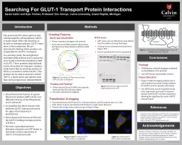
Introduction
Methods
Conclusions
References
The protein GLUT1 allows glucose into cells through the cell membrane. GLUT1 is made inside of the cell; however, to be able to transport glucose, GLUT1 must move to the membrane. We are interested in finding which proteins are responsible for GLUT1’s transport.
In a previous study, the endoplasmic reticulum (ER) proteins sec23 and sec24 were found to have the potential to bind to GLUT1. These proteins help facilitate vesicle formation for transport. Another study found that an envelope protein of HTLV-2 can bind to surface GLUT1. This protein can be used to measure surface GLUT1, a much easier and quicker task than normal expression
measurement.
Clone fluorescent fusions of a green fluorescent protein (GFP) vector and different versions of the ER proteins sec23 and sec24
Co-transfect the above fusions with mCherry_GLUT1 fusions and search for signs of interaction (co-localization)
Clone fluorescent fusions of GFP and the GLUT1-binding envelope protein of HTLV-2
Recreate a procedure given in literature using the env-GFP fusion to determine surface expression of GLUT1 in transfected cells
Sec23 and Sec24 DNAPrimer sequences designed and orderedsec23 and sec24 DNA isolated with primers by Polymerase Chain Reaction (PCR) on HK2 cell DNA
Al-Saleem J, Dirksen WP, Martinez MP, Shkriabai N, Kvaratskhelia M, Ratner L, et al. (2019) HTLV-1 Tax-1 interacts with SNX27 to regulate cellular localization of the HTLV-1 receptor molecule, GLUT1. PLoS ONE 14(3): e0214059. https://doi.org/10.1371/journal.pone.0214059
Findings Preliminary confocal imaging confirmed co-localization to be presentenv-GFP fusion successfully createdFuture DirectionLarger confocal imaging sample size is now needed to provide strong evidence for GLUT1 and sec protein interactionenv-GFP fusion can be transfected into cells, expressed, and used to measure surface level GLUT1 via flow cytometry, allowing for significantly easier GLUT1 measurement
Figures 4 and 5: mCherryGLUT1 GFP_sec24a co-transfection
Objectives
Searching For GLUT-1 Transport Protein Interactions
Sarah Catlin and Kyle Timmer, Professor Eric Arnoys. Calvin University, Grand Rapids, Michigan
Acknowledgements
Thanks to Professors Westrate, Looyenga and Baker for their guidance, Hope College for the use of their facilities, the Calvin Research Fellowship program for student funding, and the Calvin Biochemistry Department for the facilities, programming, and ice cream!
10848-1
calvingraphics@gmail.com
Transfection & ImagingFlorescent fusions of GFP_[sec23 or 24] and mCherry_GLUT2 inserted into cos7 cellsCells “fixed” in place and stained with Hochest to make nuclei fluoresceViewed cells using confocal microscope at Hope College
Figure 2:
Gene Map of a Fluorescent Fusion (GFP_sec23b) via SnapGene
HTLV-2 env
GFP vector and env DNA (from Gene Block) fused via Gibson Cloning TechniqueCloning success checked with Restriction Digest, PCRFusion transfected to test for expression
Figure 3: Restriction Digest and PCR Confirmation of env_GFP
Figure 1
: GLUT1 Structure (Adapted from
Suls et. Al (2008). Brain 131. 1831-44.
Creating Fusions
“Cutting and Pasting”
Vector and sec23/sec24 DNA cut at specific, identical sites by restriction digestCut ends reattached via ligation
Figure 6
: mCherryGLUT1 GFP_sec24b co-transfection