/
Oropharynx  surgery Assis Oropharynx  surgery Assis

Oropharynx surgery Assis - PowerPoint Presentation

DancingDragonfly
DancingDragonfly . @DancingDragonfly
Follow
342 views
Uploaded On 2022-08-04

Oropharynx surgery Assis - PPT Presentation

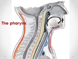
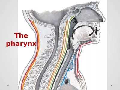
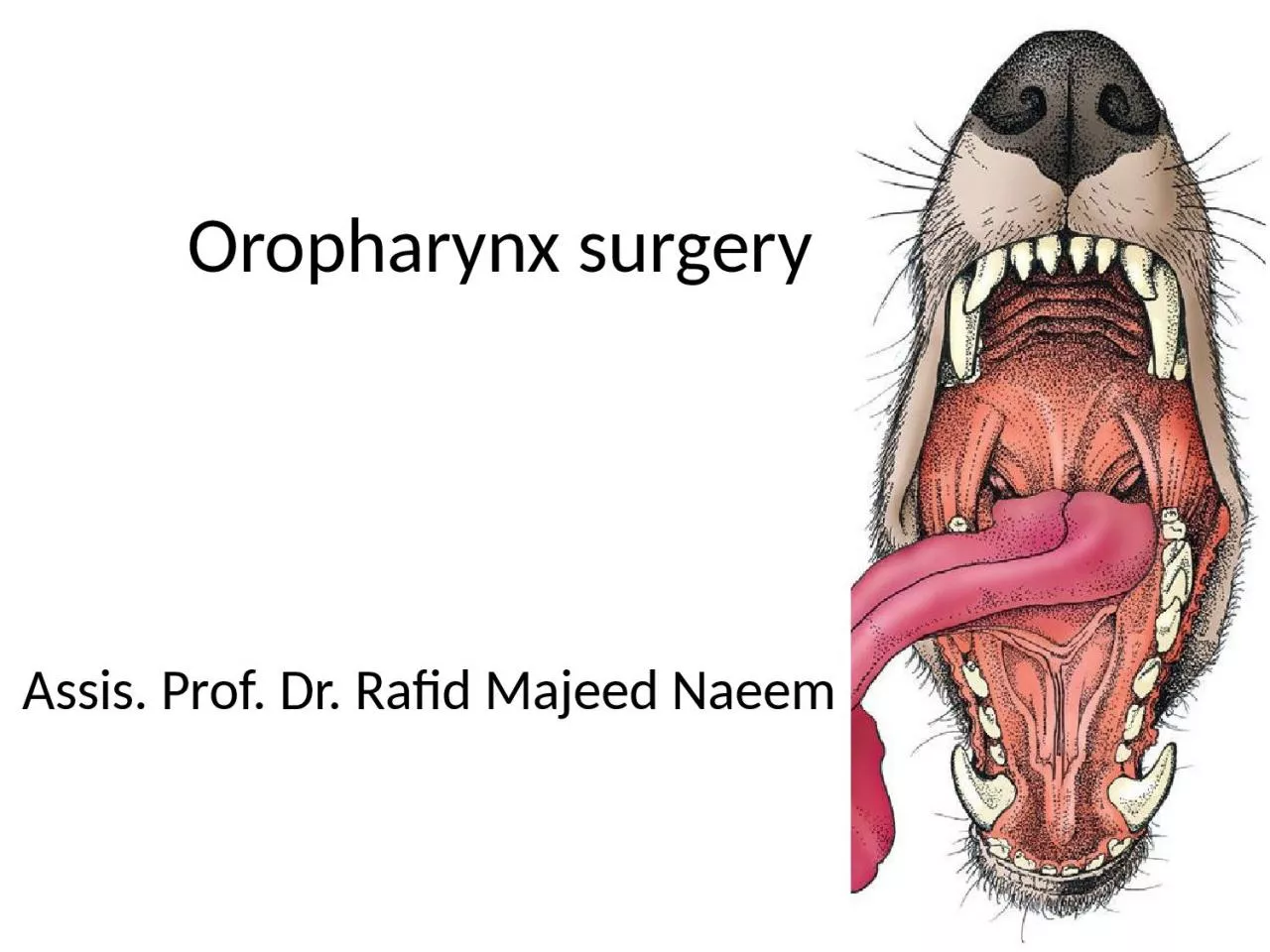
Prof Dr Rafid Majeed Naeem Tongue surgery The tongue is the most versatile organ in the oral cavity It is responsible for food prehension water lapping sucking mastication tasting swallowing grooming thermoregulation and vocalization ID: 935098

oral tongue food pharyngeal tongue oral pharyngeal food dysphagia pharynx dogs stage lingual swallowing common muscles deglutition mouth injury

Share:

Link:

Embed:

Download Presentation from below link

Download Presentation The PPT/PDF document "Oropharynx surgery Assis" is the property of its rightful owner. Permission is granted to download and print the materials on this web site for personal, non-commercial use only, and to display it on your personal computer provided you do not modify the materials and that you retain all copyright notices contained in the materials. By downloading content from our website, you accept the terms of this agreement.


Presentation Transcript

Slide1

Oropharynx surgery

Assis

. Prof. Dr.

Rafid

Majeed

Naeem

Slide2

Tongue surgery

The tongue is the most versatile organ in the oral cavity. It is responsible for food prehension

, water lapping, sucking, mastication, tasting, swallowing, grooming, thermoregulation, and vocalization.

Most of these functions require precise motor control, which is why the tongue consists almost entirely of skeletal muscle

Slide3

The tongue consists of

1. a root, which anchors it to the oropharynx;

2. a

body, which extends

rostral

to the root and is attached to the floor of the oral cavity via the frenulum; and 3. the apex, which is

rostral

and unattached to the frenulum.Adjacent to each side of the frenulum is a raised area of mucosa running longitudinally called the sublingual fold.  

This fold courses

rostrally, ending at the sublingual caruncle. The mandibular and sublingual ducts course under the mucosal folds and open at the caruncle

Slide4

The root of the tongue consists of a set of three paired extrinsic muscles:

the styloglossus,

hyoglossus

, and

Genioglossus

acts to retract and depress the tongue depress and protrude the tongue. The intrinsic muscles of the tongue are complex and often course into one another with no discernible borders

.

are responsible for protruding the tongue and a variety of other intricate movements.

Slide5

The

lyssa is a tubelike structure of muscle, fat, and sometimes cartilage encased in a dense sheath of connective tissue that lies on the median plane on the ventral aspect of the tongue. The function of the

lyssa

is unclear; however, one theory is that it serves as a stretch receptor for the tongue

Slide6

The dorsal surface of the tongue is covered with a thick mucous membrane consisting of

cornified squamous epithelium. Within the mucous membrane are structural units called papillae

that

help protect the tongue from objects that enter the mouth and assist in

prehension of food and water.

Slide7

Fungiform

,

vallate

, and foliate papillae are gustatory and therefore contain taste buds.

Nongustatory

papillae include

filiform

and conical papillae.

الحليمات الفطرية

والفايتية والورقية هي الحليمات الذوقية وبالتالي تحتوي على براعم التذوق. وتشمل الحليمات غير المستقرة الحليمات الخيطية والمخروطية الشكل.Conical

papillae have the added function of facilitating grooming by acting as a type of comb, especially in the feline tongue, where the papillae are shaped like hooks. A thin line of hair growth in the median

sulcus

(

hairy tongue

) can be a normal finding in some dogs.الحليمات المخروطية لها وظيفة إضافية تتمثل في تسهيل الاستمالة من خلال العمل كنوع من المشط ، خاصة في لسان القطط ، حيث تتشكل الحليمات مثل الخطافات. يمكن أن يكون نمو الشعر في خط رفيع في التلم المتوسط ("اللسان المشعر") نتيجة طبيعية في بعض الكلاب.

Slide8

The ventral surface of the tongue is covered in a thin mucous membrane, through which the lingual vein can often be seen coursing longitudinally on each side of the

frenulum. The lingual artery can be palpated alongside the lingual vein. The right and left lingual arteries

anastomose

throughout parenchyma of the tongue muscle; therefore, disruption of blood flow through one artery has no significant effect on blood flow to lingual tissue.

The lingual artery is a branch of the internal carotid artery and is the principle source of blood to the tongue. The lingual vein starts at the apex of the tongue, courses alongside the lingual artery, and eventually empties into the facial vein.

Slide9

DISORDERS OF THE TONGUE

Congenital

Disorders

Tongue malformations such as

macroglossia,

microglossia, and tongue deviations are rare in dogs and cats. Ankyloglossia

is a congenital disorder in which the lingual

frenulum is abnormally short and thickened, restricting movement. Affected dogs have difficulty suckling, licking, swallowing, and vocalizing, resulting in stunted growth, ptyalism, and difficulty eating. The condition has only been described in Anatolian shepherd dogs

Slide10

Infectious Disorders

The

tongue is a rare site for infection, probably because of its rich blood supply, ability to avoid penetrating injury, tough dorsal surface, and continual contact with saliva, which has antibacterial properties.

Lingual abscesses in dogs have been described and are thought to result from penetrating injury through the oral mucosa. The most common site of penetrating oral injury is directly underneath the tongue

.

Treatment consists of passive or active drainage.

Marsupialization

of the abscess into the oral cavity after surgical drainage may help prevent reformation of the abscess and provides a route for drainage

Slide11

Miscellaneous Conditions

a.

Calcinosis

circumscripta is an uncommon syndrome of ectopic mineralization that has been described in dogs, cats, and other domestic and wild mammals. The mineralization consists of calcium

hydroxyapatite crystals or amorphous calcium phosphate

and is thought to occur as a result of tissue injury (dystrophic), abnormal calcium or phosphate metabolism secondary to renal failure (metastatic), or unknown etiology (idiopathic).

The cause of the dystrophic mineralization is hypothesized to be repetitive tissue injury. Surgical excision may be performed; however, treatment is usually not necessary unless dysphagia is present.

Slide12

Oral

papillomatosis usually affect dogs younger than 1 year of age and has a viral cause. White, translucent nodules on the tongue and gingiva may be quite numerous. Lesions regress without any specific treatment within 4 to 8 weeks

.

Adult dogs with

papilloma lesions should be screened for immunosuppressive

conditionsTreatment with azithromycin62 and recombinant vaccine32 has been described in small numbers of dogs. Surgical excision of the lesions is only indicated for diagnostic and palliative purposes because the lesions tend to recur after excision

Miscellaneous

Conditions

Slide13

Trauma

Penetrating trauma

to the mouth from foreign objects is sometimes seen in dogs that chew on or carry sticks and is seen rarely in cats. In one study, the most common site of injury was sublingual.


Other reported forms of trauma include

injury secondary to tongue entrapment in a paper shredder, resulting in multiple lacerations and traumatic amputation of the entire tongue body;

piranha bite; and

entrapment of the tongue in a cage grate and a chain link fence. Burns from electrical cords are sometimes seen in young dogs and cats and often involve the hard palate and gingiva.

 Lacerations should be debrided and lavaged; most can be closed primarily. Normal tongue conformation is maintained by accurate apposition of deep muscular tissue and then epithelium with fine, rapidly absorbable, synthetic monofilament suture (3-0 to 4-0). Dogs will usually eat immediately after surgery; cats’ appetites are less predictable. Partial

glossectomy and glossectomy are discussed in the following section.

Slide14

Neoplasia and

Hyperplastic Lesions

The

most common malignant oral tumors in dogs are malignant melanoma and

squamous

cell carcinoma followed by fibrosarcoma. Gingiva

is the most common site for oral tumors in the dog followed by the tonsils and lips.

Squamous cell carcinoma accounts for approximately 70% to 80% of all oral tumors in cats followed by fibrosarcoma.The gingiva and tongue are the most frequent locations for oral tumors in the cat.14 

Tumors of the tongue make up a small percentage (3% to 4%) of all oropharyngeal tumors in the dog.  Malignant melanoma and

squamous cell carcinoma are the most common lingual tumors followed by squamous papilloma, plasmacytoma, and fibrosarcoma.

Slide15

The

oral cavity proper

refers to the space between the lower and upper dental arcades.

Oropharynx

:

The space ventral to the soft palate bound caudally by the pharyngeal wall.

The

oropharynx

is the space between the oral cavity proper and the

laryngopharynx and nasopharynx.It is bound dorsally by the soft palate and ventrally by the root of the tongue.

Slide16

The soft palate helps protect the nasopharynx

from entrance of food during deglutition by becoming taut and elevated by tensor and levatorvelipalatini muscles, respectively.  This action results in the caudal free edge of the soft palate pressing on the pharyngeal wall, sealing off the

nasopharynx

.

Slide17

Physiology of Deglutition

Swallowing, also called deglutition, is the transport of food or water from the mouth to the stomach and is one of the most complicated actions controlled by the central nervous system. It requires the coordinated actions

حركات منسقة

of muscles in the mouth, pharynx, larynx, and esophagus, all of which

are under the control of cranial nerves and the swallowing center located in the brainstem

مركز البلع في جذع الدماغ . Alterations in this process can cause dysphagia عسر البلع, aspiration, or regurgitation.


The entire process of deglutition has been divided into three phases: oropharyngeal, esophageal, and gastroesophageal.

Slide18

T

he oropharyngeal phase has been further subdivided into the oral, pharyngeal, and

pharyngoesophageal

stages

.

The oral stage involves formation of a food bolus by compression of food between the tongue and palate. The bolus is then propelled toward the base of the tongue at the entrance of the pharynx.

This stage is completely voluntary, with sensory and motor innervation to the muscles of mastication, soft palate, and tongue provided by CNs V Trigeminal nerve., VII Facial nerve., and XII Hypoglossal.

2. The bolus at the base of the tongue stimulates the reflex pharyngeal stage of deglutition. The tongue and pharyngeal constrictor muscles transport the food bolus through the pharynx through a peristaltic-like manner.

To prevent food from entering the airway, the epiglottis covers the glottis, and the soft palate presses against the pharyngeal wall to block the entrance to the nasopharynx.

Slide19

The pharyngeal stage is involuntary and under the control of the swallowing center and CNs IX Glossopharyngeal and X

Vagus . 3. The passage of food from the pharynx through the cricopharyngeal

sphincter and into the esophagus is

the

pharyngoesophageal

stage of deglutition.Relaxation of the sphincter starts as soon as the pharyngeal muscles begin constriction. The degree of sphincter opening varies with the size and consistency of the bolus.

The

pharyngoesophageal stage terminates with closure of the sphincter and relaxation of the pharyngeal constrictor muscles. This stage is also involuntary and under the control of CNs IX and X.

Slide20

GENERAL CONSIDERATIONS

Clinical Signs and DiagnosticsGeneral clinical signs of oral disease include ptyalism

(an abnormal and excessive secretion of saliva.),

dysphagia

(a difficulty in swallowing ),inappetence (reduction or complete loss of appetite),

weight loss

, pain, halitosis (Bad mouth smell), and oral hemorrhage

.

Slide21

In patients known or suspected to have

oral neoplasia, cytologic evaluation of fine needle aspirates taken from

mandibular

lymph nodes is recommended

.  

Medial retropharyngeal lymph nodes are palpable only when they are significantly enlarged.Evaluation of a three-projection

thoracic radiograph study

is a good for detection of metastasis in the lungs and other thoracic structures or if lower airway tract disease is suspected.

Slide22

Preoperative Preparation

Tumors or swelling in the oral or pharyngeal cavity can complicate intubation.A laryngoscope with adequate lighting,

endotracheal

tubes of multiple sizes,

and availability of an oxygen mask are essential.

When anesthetizing patients with large masses or for those that have already displayed signs of dyspnea

, instruments for performing an

emergency tracheotomy should be available. 

Slide23

After the endotracheal tube is in place, it should be secured so as to not interfere with the surgical site

An appropriately sized endotracheal tube (with a properly inflated cuff),

along with absorbent material packed in the back of the pharynx, will usually prevent significant leakage into the trachea.

Slide24

Visualizing the pharynx and

evacuating any fluid with a suction tube immediately before extubation may also help prevent fluid aspiration and will ensure that packing material is not left in the pharynx..


Povidone

– iodine solution or a 0.2% chlorhexidine

solution is used for preparation of the oral mucosa. Perioperative antibiotics are generally not indicated because the mouth is remarkably resistant to infection, presumably because of its excellent blood supply and the antibacterial properties of saliva.

Slide25

DISORDERS OF THE OROPHARYNX

DysphagiaDysphagia:

is difficulty swallowing during any stage of deglutition


Symptoms of dysphagia affecting the oral stage

of deglutition may include difficulty prehending food or lapping water; retention of food or water in the mouth; and failure to swallow saliva, resulting in drooling.

Some animals are able to compensate by making sudden head movements while eating in an attempt to move food back into the pharynx.

If other stages of deglutition are normal, animals with oral stage dysphagia may benefit from feeding and providing water in an elevated position to aid delivery of food and water to entrance to the pharynx.

Slide26

Food may be regurgitated back into the oral cavity or may stay in the pharynx.

Because the epiglottis relaxes at the termination of swallowing and leaves the airway unprotected, affected animals are at risk for laryngotracheal

aspiration.

In animals with dysphagia of the pharyngeal or

pharyngoesophageal

stage, function is not improved by feeding in an elevated position or placing food at the base of the tongue.

Clinical features of dysphagia during the pharyngeal or

pharyngoesophageal stages usually include

repeated unsuccessful attempts at swallowing.

Slide27

Etiology and Diagnosis

Dysphagia usually results from space-occupying masses, structural congenital abnormalities, or functional abnormalities.

Structural Abnormalities and Masses.

Structural congenital abnormalities resulting in

dysphagia include congenital palate defects,

ankyloglossia (limited normal movement of the tongue chiefly due to an abnormally shortened frenulum : tongue-tie),

macro- and

microglossia, and tight lip syndrome (reduced mandibular vestibule). These should be easily detected on oral examination under sedation or anesthesia. Space-occupying masses resulting in dysphagia

include extrapharyngeal masses, such as enlarged retropharyngeal node(s), cervical abscess or granuloma (a focal aggregate of immune cells), and invasive salivary or thyroid neoplasms

, and masses of the palatine tonsils.

Slide28

Treatment depends on the specific cause. A mass in the oral cavity may not cause any symptoms until it reaches a critical size, subsequently resulting in drooling, difficulty in food

prehension, or malodor. Masses located in the pharynx usually result in difficulty swallowing or

dyspnea

and require sedation or anesthesia to visualize except in the most cooperative patients.

Pharyngeal masses that result in an acute onset of

dyspnea require rapid decompression (e.g., incisional drainage of mucoceles

) or intubation to quickly

unobstruct the airway. Dysphagia from penetrating injuries may occur from the presence of a foreign body or swelling, pain, or infection associated with the trauma.

Slide29

Fluoroscopic evaluation of the swallowing process using barium contrast is the most useful tool in diagnosing and further characterizing functional

dysphagia. Patients with pharyngeal

dysphagia

will make repeated attempts to swallow, but the food bolus fails to move past the pharyngeal constrictor muscles and remains in the pharynx.

In contrast, patients with

pharyngoesophageal dysphagia will have normal movement of the bolus through the pharynx, but the bolus will fail to pass through the

cricopharyngeal

sphincter, either remaining within the pharynx or redirecting into the larynx. The most common pharyngoesophageal dysphagias are cricopharyngeal asynchrony and cricopharyngeal

achalasia.

Slide30

Penetrating Injuries to the

OropharynxPenetrating trauma to the mouth from foreign objects is sometimes seen in dogs that carry or chew sticks and is seen rarely in cats.


The most common presenting complaints are

dysphagia, drooling, depression,

oral pain, pain on flexion of the neck, subcutaneous emphysema in the cervical region, blood in the saliva, and pain on opening of the mouth.

Cervical, cranial, or facial swelling is a common presenting complaint in chronic cases.

Additionally, chronic cases may have a draining sinus tract in the cervical region.

Slide31

Diagnostics

Plain radiographs of the cervical region, including the retropharyngeal space, are recommended for initial diagnostic workup in animals with a suspected or confirmed penetrating injury to the oropharynx or esophagus.

Slide32

Common sites of penetration are the esophagus, lateral and dorsal pharyngeal walls, and sublingual region.

The decision to use advanced imaging is based on the chronicity of the injury, the presence of cervical swelling, and radiographic signs of gas or tissue reaction within the cervical region.

Slide33

Surgery

Wound exploration, debridement, and lavage may be adequate treatment in most cases of penetrating trauma without extraluminal pharyngeal involvement.

Surgical exploration of the retropharyngeal space is accomplished through a ventral midline approach.

Care is taken to avoid disruption of the recurrent laryngeal nerves during dissection.


The goal is to locate and remove foreign material, obtain tissue or fluid samples for cytology and culture,

debride

nonviable tissue, and establish drainage if necessary.

Slide34

administration of antibiotics is usually indicated if infection is suspected.

Therapy can be modified after culture and sensitivity results are available.