

91 Joints and their classifications Joint any point where two bones meet both movable and immovable Arthrology science of joint structure function and dysfunction Kinesiology study of musculoskeletal movement ID: 934917
Download Presentation The PPT/PDF document "joints Chapter 9 9.1 Joints and their Cl..." is the property of its rightful owner. Permission is granted to download and print the materials on this web site for personal, non-commercial use only, and to display it on your personal computer provided you do not modify the materials and that you retain all copyright notices contained in the materials. By downloading content from our website, you accept the terms of this agreement.


Slide1
joints
Chapter 9
Slide29.1 Joints and their Classifications
Slide39.1 Joints and their classifications
Joint
:
any point where two bones meet; both movable and immovable
Arthrology
: science of joint structure, function and dysfunctionKinesiology: study of musculoskeletal movementBranch of biomechanics, which deals with body movements in general
Slide49.1 Joints and their classifications
Names of joints are typically derived from the names of the bones involved…
Atlantooccipital
joint
Glenohumeral
jointRadioulnar jointEtc.
Slide59.1 Joints and their classifications
Joints are classified according to the manner in which the adjacent bones are bound to each other
Bony
Fibrous
Cartilaginous
Synovial
Slide69.1 Joints and their classifications
Joint Classification
Bony
– (
synostosis
) immovable joint formed when the gap b/t two bones ossifies and they become a single bone
Slide79.1 Joints and their classifications
Joint Classification
Fibrous
– (
synarthrosis
) adjacent bones are bound by collagen fibers
Slide89.1 Joints and their classifications
Fibrous
Sutures:
immobile or only slightly moveable; closely bind skull bones to each other
Gomphoses
: attachment of a tooth to its socketSyndesmoses: fibrous joint at which two bones are bound by relatively long collagenous fibers; radius to ulna
Slide99.1 Joints and their classifications
Joint Classification
Cartilaginous
– (
amphiarthrosis
) two bones are linked by cartilage
Slide109.1 Joints and their classifications
Cartilaginous
Synchondroses
:
joint in which bones are bound by
hyaline cartilage; growth plates, first rib attachment to sternumSymphyses: two bones are joined by
fibrocartilage
; pubic symphysis, between two vertebrae
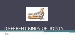
Slide119.2 Synovial joints
Slide129.2 Synovial Joints
Synovial joints
: (
diarthrosis)
Most familiar type
Most freely movableMost structurally complexMost likely to develop uncomfortable and crippling dysfunctions
Slide139.2 Synovial Joints
Structural Anatomy
Articular cartilage
: layer of hyaline cartilage on the facing ends of two bones
Joint cavity
: narrow space separating bone surfacesSynovial fluid: Slippery lubricant which nourishes the cartilage
Slide149.2 Synovial Joints
Structural Anatomy
Joint capsule
: connective tissue which encloses cavity and retains fluid
Fibrous capsule (outer)
: continuous with periosteum of adjoining bonesSynovial membrane (inner): secretes synovial fluid
Slide159.2 Synovial Joints
Structural Anatomy
In a few synovial joints, fibrocartilage grows inward from joint capsule, forming a pad in b/t bones
Jaw, radius/ulna, ulna and carpal bones =
articular disc
Knee = meniscus
Slide169.2 Synovial Joints
Structural Anatomy
Accessory structures
are also associated with synovial joints:
Tendon
- strip/sheet of touch collagenous CT that attaches muscle to bone (tendon sheaths)Ligament – attaches one bone to anotherBursa – fibrous sac filled with synovial fluid; located b/t two muscles
Slide17Exercise and Articular Cartilage
When synovial fluid is warmed by exercise, it becomes thinner and more easily absorbed by the articular cartilage.
Repetitive compression of the cartilage, during exercise, is important to its nutrition and waste removal.
Swimming
is one of the best ways of exercising the joints with minimal damage
Slide189.2 Synovial Joints
Terms associated with levers…
Effort arm
: portion o f lever from fulcrum to point of effort (muscle)
Resistance arm
: portion of lever from fulcrum to point of resistance (object or body itself)Fulcrum: (joint)Joints and Lever SystemsMany bones, especially the long bones, act as
levers
to enhance the speed or power of limb movements
Slide199.2 Synovial Joints
1
st
class lever
– teeter-totter/muscles of the back of the neck pulling on occipital bone
2nd class lever – wheel barrow/quads of thigh elevating the knee3rd class lever – canoe paddle/ biceps flexing the elbow
Slide209.2 Synovial Joints
Normally determined by…
Structures of articulating surfaces
Strength and tautness of ligaments and joint capsules
Action of the muscles and tendons
Range of Motion (ROM): degrees through which a joint can move
Slide219.2 Synovial Joints
Multiaxial
– three degrees of rotation (shoulder)
Biaxial
– two degrees of rotation (metacarpophalangeal)
Monoaxial- one degree of rotation (elbow)Axes of Rotation: pass through bone in a direction perpendicular to the plan of movement
Slide229.2 Synovial Joints
Ball-and-socket
– smooth hemispherical head fitting into a cuplike socket
Condylar
- oval convex surface fitting into a complementary shaped depression
6 class of jointsDistinguished by
shapes
of surfaces and
degrees of freedom
Slide239.2 Synovial Joints
Saddle
– both bones have a saddle shaped surface – concave in one direction
Plane
– Surfaces are flat or only slightly curved
6 class of joints
Slide249.2 Synovial Joints
Hinge
– cylindrical surface which fits into corresponding depression
Pivot
– bone spins on its longitudinal axis
6 class of joints
Slide259.2 Synovial Joints
Movements of Synovial Joints
Kinesiology, physical therapy and other medical and scientific fields have a specific vocab. for movements of synovial joints
All return to
zero position
– position when person is viewing in anatomical position
Slide269.2 Synovial Joints
Movements of Synovial Joints
Flexion
: mvmt that decreases a joint angle
Extension
: mvmt that straightens a joint; returns to 0
Slide279.2 Synovial Joints
Movements of Synovial Joints
Hyperextension
: extension of the joint beyond the 0 position
Ligaments or bone structure typically prevents this.
Slide289.2 Synovial Joints
Movements of Synovial Joints
Abduction
: mvmt away from the midline of the body
Adduction
: mvmt back toward the midline
Slide299.2 Synovial Joints
Movements of Synovial Joints
Elevation
: mvmt that raises a body part
Depression:
mvmt that lowers a body part
Slide309.2 Synovial Joints
Movements of Synovial Joints
Protraction
: mvmt of a body part in the anterior
Retraction:
mvmt of body part in posterior direction
Slide319.2 Synovial Joints
Movements of Synovial Joints
Circumduction
: One end of an
a
ppendage remains fairly stationary while other makes a circular motionRotation: Mvmt in which bone spins on long. axis
Slide329.2 Synovial Joints
Movements of Synovial Joints –
Primarily forearm movements
Supination
: Mvmt that turns pal to face up or outward
Pronation: Mvmt causing palm to face down or backwards
Slide339.2 Synovial Joints
Movements of Synovial Joints –
Special movements of the head/trunk
Lateral flexion
: tilting head or trunk to R of L of midline
Right or left rotation: twisting at waist or turning of head
Slide349.2 Synovial Joints
Movements of Synovial Joints –
Special movements of the mandible
Lateral/medial excursion
: movement to the L or R of zero position
Slide359.2 Synovial Joints
Movements of Synovial Joints –
Special movements of the hands/digits
Ulnar flexion
: tilts hand
twd little fingerRadial flexion: tilts hand twd thumb
Slide369.2 Synovial Joints
Movements of Synovial Joints –
Special movements of the hands/digits
Radial abduction
: moving thumb away from index finger (
forming an L)Palmar abduction: move thumb away from plane of hand (like wrapping hand around a tool handle)Opposition/reposition: Moving thumb to touch tip of any finger; returning to zero
Slide379.2 Synovial Joints
Movements of Synovial Joints –
Special movements of the foot
Dorsiflexion
: mvmt in which toes are elevated
Plantar flexion: mvmt in which toes point downwardsInversion/Eversion: Tilting soles medially or laterally (respectively)
Slide389.3 Anatomy of Selected Diarthroses
Slide399.3 anatomy Selected diarthroses
The joints discussed in this section are the most likely to require medical attention and have strong bearing on athletic performance and everyday mobility
Slide409.3 anatomy Selected diarthroses
Jaw joint:
Temporomandibular joint (TMJ)
Articulation of the condyle of the mandible with the mandibular fossa of the temporal bone
Lateral ligament
– prevents posterior displacementTMJ Syndrome
Slide419.3 anatomy Selected diarthroses
Shoulder joint:
Glenohumeral
(humeroscapular) joint
Articulation of the
humerus and the glenoid cavity of the scapulaRotator cuff: several tendons which fuse to the joint capsule to stabilize the jointShoulder dislocation
Slide429.3 anatomy Selected diarthroses
Elbow joint:
Two articulations –
humeroulnar
joint and the
humeroradial jointOlecranon bursa: eases movement of tendons over jointBursitis
Slide439.3 anatomy Selected diarthroses
Hip joint:
Coxal
joint
Articulation of the head of the femur with the acetabulum of the hip bone
In a standing position, various ligaments twist to pull head of femur tightly into socketHip dysplasia
Slide449.3 anatomy Selected diarthroses
Knee joint:
Tibiofemoral joint
Largest and most complex
Mainly stabilized by quadriceps tendon in from and semimembranosus muscle in the rear
Menisci: c-shaped cartilages (lateral and medial); absorb shock of body weight
Slide459.3 anatomy Selected diarthroses
Knee joint:
Collateral ligaments
(
lateral and medial
) prevent knee from rotating when joint is extendedCruciate ligaments (anterior and posterior) prevent hyperextension and prevent the femur from sliding off the front of the tibia
Slide469.3 anatomy Selected diarthroses
Ankle Joint:
Talocrural
joint
Two articulations – tibia to talus and fibula to talus
Malleoli – knobs of the ankles; restricts ROMCalcaneal tendon – extends from calf muscles to calcaneus and allows for dorsiflexion and plantar flexion
Slide479.3 anatomy Selected diarthroses
Table 9.1 on page 306
lists a number of common joint disorders and the various clinical applications in this chapter cover other joint disorder/injury related topics
Slide48Chapter 9 Review:
www.studystack.com/menu-845748