
SR JNMC ALIGARH This is stalklike part of the brain which connects the forebrain with the spinal cord It consists from below upward of the medulla oblongata pons and midbrain ID: 930906
Download Presentation The PPT/PDF document "MEDULLA BY Dr ROBERTON GAUTAM" is the property of its rightful owner. Permission is granted to download and print the materials on this web site for personal, non-commercial use only, and to display it on your personal computer provided you do not modify the materials and that you retain all copyright notices contained in the materials. By downloading content from our website, you accept the terms of this agreement.


Slide1
MEDULLA
BY Dr ROBERTON GAUTAM
SR, JNMC ALIGARH
Slide2This is stalk-like
part of the brain which
connects the forebrain with the spinal cord. It consists, from below upward, of the medulla oblongata, pons, and midbrain.The brainstem consists of nerve fibres and nerve cells.The brainstem nuclei are of the following two types:1. Nuclei of last 10 cranial nerves (i.e., 3rd–12th cranial nerves).2. Other named nuclei such as red nucleus, substantia nigra, pontine nuclei, olivary nuclei, etc.In addition to well-defined tracts and nuclei, the brainstem consists of the diffuse system of cells and fibres called reticular formation. Some of the cells of reticular formation form vital centres, viz. cardiac, respiratory, vasomotor, etc.
Brainstem
Slide3The
medulla oblongata is the direct upward continuation
of the spinal cord, extending from the foramen magnum to the upper border of the pons. It forms the lowest part of the brainstem and lies almost vertically in the anterior part of the posterior cranial fossa between the clivus in front and the vallecula of the cerebellum behind.Medulla provides attachment to the last four cranial nerves.MEDULLA OBLONGATA
Slide4The lower part of the medulla, like the spinal cord, contains the central canal.
In the upper part of the medulla, this canal widens and moves dorsally to form the lower part of the 4th ventricle.
Thus, the medulla is divided into two parts: a lower closed part of medulla and an upper open part.
Slide5Features on the Ventral
Aspect
The medulla is divided into right and left symmetrical halves - by the anterior median fissure and posterior median sulcus.Anterior median sulcus is interrupted in its lower part by the bundles of fibres crossing obliquely from one side to the other, the decussation of pyramids.The posterior median sulcus is present only in the lower half of the medulla. Above, its lips diverge to form the boundaries of a triangular area, the lower part of the floor of the 4th ventricle.
Slide6Features on the Ventral
Aspect
Each half of the medulla is marked by two sulci, anterolateral and posterolateral, which are direct upward continuations of the corresponding sulci of the cord.The anterolateral sulcus extends along the lateral border of the pyramid and along it emerge the rootlets of the XII Cranial nerve.The posterolateral sulcus lies between the olive and the inferior cerebellar
peduncle and along it emerge the rootlets of
the IX, X,
and
XI
cranial nerves.
Slide7The
ventral aspect of medulla presents the
following features:1. Pyramids.2. Olives3. Rootlets of the hypoglossal nerve.4. Inferior cerebellar peduncles5. Rootlets of the IX, X, and XI (cranial part) cranial nerves
Slide8The
closed part, on either side of the posterior
median sulcus, presents three longitudinal elevations. From medial to lateral these are: fasciculus gracilis,fasciculus cuneatus and inferior cerebellar peduncle.The upper ends of the fasciculus gracilis and fasciculus cuneatus expand to form the gracile and cuneate tubercles, respectively.Another elevation present lateral to cuneate tubercle; the tuber cinereum is produced by the spinal nucleus of the trigeminal nerve.
Features of the Closed
Part
Slide9The open part of the medulla forms
- the
lower part of the floor of the 4th ventricle.Features of the Open Part
Slide10It is studied in transverse sections (T.S.) at the
three levels
: 1. At the level of decussation of pyramids. 2. At the level of sensory decussation. 3. At the level of the olives.INTERNAL STRUCTURE
Slide11Features on the Dorsal
Aspect
The dorsal aspect of the medulla is well demarcated into lower closed and upper open parts.
Slide12Transverse section of medulla at the level
of pyramidal
decussation.The transaction at this level passes through the inferior half of the medulla.1. The nucleus gracilis and nucleus cuneatus 2. Nucleus of the spinal tract of the trigeminal nerve.3. The spinal tract of the trigeminal nerve 4. Decussation of pyramidal tracts forms the mostimportant feature of medulla at this level. 5. Each detached anterior horn divides to form the spinal nucleus of the accessory nerve and the supraspinal nucleus of the 1st cervical nerve.6. Diffuse zone appears containing a network of fibres and scattered nerve cells within it
.
Slide131
. The
nucleus gracilis and nucleus cuneatus become more pronounced with their fibres.2. The internal arcuate fibres arising from gracile and cuneate nuclei.3. The internal arcuate fibres cut off the spinal nucleus and tract of the trigeminal nerve.4. Accessory cuneate nucleus 5. The separated spinal nucleus and tract of trigeminal Nerve.6. The lower part of inferior olivary nucleus .7. The pyramids
8. The central grey matter contains the: (a)
hypoglossal nucleus, (b) dorsal nucleus of
vagus
, and (c) nucleus of
tractus
solitarius
.
9. The
medial longitudinal bundle/fasciculus
10.
Spinocerebellar
and lateral
spinothalamic
tracts.
11.
Lateral and anterior
spinothalamic
tracts
collectively form
spinal
lemniscus.
T.S. of Medulla at the Level of Sensory
Decussation
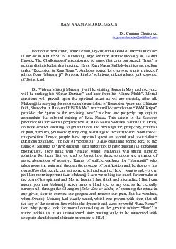
Slide14Transverse
section of medulla at the level
of sensory decussation (1 = nucleus tractus solitarius, 2 = dorsal nucleus of vagus, 3 = hypoglossal nucleus; M = medial longitudinal fasciculus, R = reticular formation).
Slide151
.
Hypoglossal nucleus, nucleus intercalatus, dorsal nucleus of vagus,and vestibular nuclei (inferior and medial).2. The nucleus of tractus solitarius lies ventral to vestibular nuclei.3. The nucleus ambiguus.4. On either side of the midline (paramedian region) from dorsal to ventral lie medial longitudinal fasciculus (MLF), tectospinal, medial lemniscus, and pyramidal (corticospinal) tracts.5. The
arcuate
nuclei
6
. Laterally
,
(a)
inferior
cerebellar
peduncle and
(
b)
inferior
olivary
nucleus
.
T.S. of Medulla at the Level of Olives
Slide16Transverse section of medulla at the level of olives
(M = medial longitudinal bundle, R =
reticular formation, T = tectospinal tract).
Slide17The medulla is supplied by the following arteries:
1. Two vertebral arteries.
2. Anterior and posterior spinal arteries.3. Anterior and posterior inferior cerebellar arteries.4. Basilar artery.ARTERIAL SUPPLY OF THE MEDULLA
Slide18CLINICAL
Lateral
medullary (posterior inferior cerebellar artery) syndrome of Wallenberg: It occurs due tothrombosis the of posterior inferior cerebellar artery.affecting a wedge-shaped area on the dorsolateral aspect of the medulla and the inferior surface of the cerebellum, and produces the signs and symptoms:– Contralateral loss of pain and temperature sensation in the trunk and limbs, due to involvement of spinothalamic tract.– Ipsilateral loss of pain and temperature sensation over the face, due to involvement of the spinal nucleus and tract of the trigeminal nerve.–
Ipsilateral
paralysis of muscles of palate, pharynx,
and
arynx
, due to involvement of nucleus
ambiguus
.
–
Ipsilateral
ataxia, due to involvement of inferior
cerebellar
peduncle
and cerebellum.
–
Giddiness, due to involvement of vestibular nuclei
.
Medial
medullary
syndrome: It occurs due to
involvement of the
paramedian
region of the medulla
following damage to penetrating branches of the anterior spinal branch of the vertebral artery.
It produces the following signs and symptoms:
–
Contralateral
hemiplegia
/paralysis of arm and leg, due
to damage of pyramid.
–
Ipsilateral
paralysis and atrophy of the half of the tongue, due to damage of hypoglossal nerve.
–
Contralateral
loss of position and vibration sense due
to damage of medial
lemniscus
.
Slide19THANK
YOU