
Nakornping hospital บาดเจบไขสนหลง คอ การบาดเจบทเกดขนกบสวนของไขสนหลง ตงแตบรเวณ ID: 934828
Download Presentation The PPT/PDF document "SPINAL CORD INJURY Rehabilitation unit," is the property of its rightful owner. Permission is granted to download and print the materials on this web site for personal, non-commercial use only, and to display it on your personal computer provided you do not modify the materials and that you retain all copyright notices contained in the materials. By downloading content from our website, you accept the terms of this agreement.


Slide1
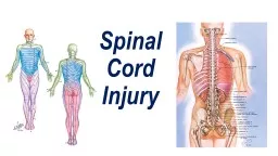
SPINAL CORD INJURY
Rehabilitation unit,
Nakornping
hospital
Slide2บาดเจ็บไขสันหลัง
คือ การบาดเจ็บที่เกิดขึ้นกับส่วนของไขสันหลัง ตั้งแต่บริเวณ
foramen magnum จนถึงส่วนปลายคือ conus medullaris ซึ่งจะอยู่ประมาณขอบล่างของกระดูก L1 หรือบนกระดูก L2 รวมทั้งส่วนของ cauda equina ด้วย
Slide3Primary Cause of Death
Slide4การตรวจทางระบบประสาทเพื่อจำแนกความรุนแรงในผู้ป่วยบาดเจ็บไขสันหลัง
American Spinal Injury Association (ASIA) guidelines
Slide5Slide6การตรวจระบบประสาทรับความรู้สึก
28 key dermatomes
ที่ต้องตรวจ pinprick & light touch ของร่างกายทั้ง 2 ซีกคะแนน 0ไม่สามารถแยกความรู้สึกแหลมกับทู่ได้คะแนน 1 แยกแหลมกับทู่ได้แต่ความรู้สึกแหลมไม่เท่ากับใบหน้าคะแนน 2 ความรู้สึกแหลมเป็นปกติเท่ากับใบหน้าการตรวจ sacral sparing ทำโดยการ PR ถ้าผู้ป่วยรู้สึกถึงการสัมผัสหรือแรงกด ถือว่ายังมี sacral sparing
Slide7Slide8การตรวจระบบประสาทสั่งการ
ตรวจกำลังของกล้ามเนื้อหลักในร่างกายทั้งสองข้าง
ตรวจผู้ป่วยในท่านอนหงายแบ่งความแข็งแรงของกล้ามเนื้อเป็น 6 ระดับ
Slide9Key Muscles
C5-elbow flexors
C6-wrist extensorsC7-elbow extensorsC8-finger flexors (distal phalanx of 3rd finger)T1-small finger abductors
Slide10ตรวจประเมิน UE
Slide11Key Muscles
L2- hip flexors
L3- knee extensorsL4- ankle dorsiflexorsL5- long toe extensorsS1- ankle plantar flexorsPR for sphincter tone assessment
Slide12ตรวจประเมิน LE
Slide13Slide14ความรุนแรงของการบาดเจ็บแบ่งตาม
ASIA impairment scale (revised 2000)
ASIA:A (complete)ASIA:B (incomplete)ASIA:CASIA:DASIA:EPrognosis
Slide15Expected Functional Outcomes by Neurologic Level of Injury
Slide16Slide17Slide18Slide19Slide20Rehabilitation Phase of Injury
Goal :
maximizing physical independencebecoming independent in direction of carePreventing secondary complicationspressure ulcerjoint stiffnessurinary tract GI tract etc.
Slide21Interdisciplinary Team
Slide22Interdisciplinary Team
The patient and family
members need to be educated about the nature of an SCIand the patient’s prognosis and the uncertainty of such
Slide23Interdisciplinary Team
Rehabilitation nurses, in addition
to performing their standard nursing duties, provideeducation on prevention and treatment of secondary complications,in addition to training in bowel and bladdermanagement.
Slide24Interdisciplinary Team
Physical and occupational therapists
in the acute hospital should facilitate prevention of secondary complications such as contractures, pressure ulcers, and disuse atrophy. This is done through maintenance of joint ROM, splinting, positioning,and selective muscle strengthening. - ROM of all joints isperformed and taught by the therapists to people with SCI and their caregivers as soon as it is medically safe to do so. - Performance of an adequate daily stretching program canprevent joint contractures. - Splinting of joints, with eitheran off-the-shelf or a custom splint fabricated by an occupational therapist, is also often used to provide a prolonged stretch, to facilitate a functional joint position, and to prevent skin breakdown.
Slide25Physical Skill Training
Mobility
self-care skillsother activities of daily living (ADL)
Slide26Practices (PT)
joint ROM and strength
Mat activities1rolling prone on elbows positioningprone on hands positioningsupine on elbows positioning,long sittingshort sittingquadruped positioning,transfer training1
Slide27Transfer trainingstand-pivot and sit-pivot transfers
Slide28Transfer trainingcomplete paraplegia/lower tetraplegia
Slide29Transfer trainingcomplete paraplegia/lower tetraplegia
1. Lift feet onto bed and wheel the chair forward against bed. Put on brakes.
Then bend forward and lift butt forward on chair.
2. With one hand on the cushion and one on the bed, lift the body sideways onto the bed.
3. Repeated lifts and lifting of legs may be needed
.
Slide30Transfer trainingThe floor-to chair transfer
1. Sit with legs straight, Pull seat to your side opposite the wheelchair (a person's knee can also be used).
2. With hands on each chair, push up, with your head forward over knees
3. Swing onto the seat.
4. Now, with your head forward over your knees, swing body onto the wheelchair.
Slide31Transfer training
The floor-to chair transfer
Slide32Standing
complete thoracic level injuries
**with caution in individuals with chronic SCI**KAFO
Slide33Wheelchair Skills
Slide34Other
Spinal Cord Injury Education
Home and Environmental ModificationsDriver TrainingVocational Training
Slide35Chronic Phase of Injury
Slide36Adjustment to Disability
Quality of Life
Recovery-Enhancing TherapiesLate Neurologic Decline
Slide37Secondary Conditions
Slide38Pulmonary System
Pulmonary complications are the leading causes of death for people with SCI
Slide39Slide40THE POSITION OF THE DIAPHRAGM
Slide41Respiratory
Level
OutcomeExpected OutcomeEquipmentC1–4VentilatorInability to clear secretionsVentilator(s) Suction equipment
Backup generator
Nebulizer
C5
C6–C7
C8
Low endurance and vital
capacity
require assist to clear secretions
-
T1–T12
Low endurance
and vital
capacity
-
L1–S5
Normal
-
Slide42Management of Pulmonary Complications
Atelectasis
pneumonia, pleural effusion, empyema
Slide43lung expansion
Intermittent positive pressure breathing
bilevel positive airway pressureContinuous positive airway pressure (CPAP)
Slide44Secretionmobilization techniques
Postural drainage
PercussionVibration
Slide45Postural drainage
Slide46Contraindication
Severe hemoptysis
Untreated acute conditionsevere pulmonary edemacongestive heart failurelarge pleural effusionPulmonary embolismpneumothorax
Slide47Contraindication
Cardiovascular instability
cardiac arrhythmiasevere hypertension or hypotensionrecent myocardial infarctionRecent neurosurgery
Slide48Precaution
Hemoptysis
PostoperationGeriatricMalignancyUnilateral lung abscess
Slide49Right and Left upper lobe
Slide50Right and Left upper lobes
Slide51Slide52Right and Left lower lobes
Slide53Right and Left lower lobes
Slide54PercussionVibration
Slide55Chest percussion
Slide56Incentive spirometer
Slide57Vascular System
Deep venous thrombosis (DVT)
Pulmonary embolism
Slide58Cardiovascular and Autonomic System
Slide59Autonomic Dysfunction
under
supraspinal controlautonomic nervous system normally controls visceral functions and maintains internal homeostasis through its nerve supply to smooth muscles, cardiac muscle, and glands
Slide60Orthostatic Hypotension
Immediately after SCI
a complete loss of sympathetic tone neurogenic (“spinal”) shock with hypotension, bradycardia, and hypothermiathe sympathetic reflex activity returns normalization of blood pressureSupraspinal control: absent in those individuals with high-level and neurologically complete SCI orthostatic hypotension
Slide61Management
elastic stockings
abdominal bindershydrationgradually progressive daily head-up tiltadministration of salt tablets, midodrine, orfludrocortisone.
Slide62Autonomic Dysreflexia
syndrome and clinical emergency that affects people with SCI usually at the T6 level or above
symptoms pounding headacheSystolic and diastolic hypertensionprofuse sweatingcutaneous vasodilatation with flushing of the face, neck,and shoulders nasal congestionpupillary dilatationbradycardia
Slide63Autonomic Dysreflexia
Triggered by a noxious stimulus below the injury level
Distended bladderfecal impactionpathology of the bladder and rectumingrown toenailslabor and deliverysurgical procedures, orgasmEtc.
Slide64Autonomic Dysreflexia
Treatment of acute AD
identification of the precipitating stimulussat upLoosen constrictive clothing and garments blood pressure monitored every 2 to 5 minutesEvacuation of the bladder doneResolved fecal impaction* Local anesthetic agents should be used during any manipulations of the urinary tract or rectum*Administered fast-acting antihypertensive agents
Slide65Bowel Management
Slide66reflexic or UMN bowel
areflexic
or LMN bowel
Slide67Upper motor neurogenic bowel (UMNB)
Suprasacral lesion
Lower motor neurogenic bowel (LMNB) Conus medullaris, Cauda equina lesion Pathophysiology of neurogenic bowel dysfunction
Slide68Upper motor
neurogenic
bowel↓Colonic motility Constipation ↓ Ability to sense the urge Loss volitional control incontinenceIntact spinal reflex (sacral)Normal or increase anal sphincter tone
Slide69Lower motor
neurogenic
bowelProlonged transit time constipation↓ anal tone incontinenceAnorectal reflex is absent or decreaseAnocutaneous reflex is absent or decrease
Slide70Summarize
LMNB
constipation with a high risk of frequent incontinence through a lax external sphincter mechanismUMNB constipation with fecal retention behind a spastic anal sphincter require a chemical or mechanical trigger for defecation
Slide71Bowel
Level
OutcomeExpected OutcomeEquipmentC1–4C5Total assist for digital stimulation, insertion of minienema or suppository, and perineal hygienePadded reclining commode chair with head support C6–C7Some to total assist for setup and perineal hygienePadded commode chairSuppository inserter
Digital bowel stimulator
Mirror
C8
T1–T12
L1–S5
Independent digital stimulation, suppository or
minienema
insertion, and perineal hygiene
Padded commode chair
Slide72Normal
Cord transection
Cauda equina
Bowel dysfn.
Normal
Constipation reflex Defecation
Constipation
Transit time
12-48 hr.
> 72 hr.
> 6 days
GMC Response to stim.
Facilitate by defecation
less
less
Slide73Anal sphincter pressure
Normal
Cord transection
Cauda equina
Resting tone
N
N
D
Volitional squeeze
N
Absent
Absent
Rectal compliance
N
N
I
Slide74Normal
Cord
transection
Cauda equina
Reflex defecation
Yes
Yes
No
Perianal sense
N
No
Loss perianal
Anal appearance
N
N
Flatten
BCR
N
Present
Absent
Anal wink
N
Present
Absent
Slide75Slide76Slide77Neurogenic
bowel management
Slide78Management
Goal of bowel program
effective and efficient colonic evacuation Bowel evacuation at a consistent time of daypreventing incontinencepreventing constipation.social continencePredictableScheduledAdequate defecation without incontinence at other time
Slide79Slide80Bowel program
Fluid
DietTimingFrequencyMedicationBowel careProcedure to periodically evacuate stool from the colon
Slide81Fluid
Must be balanced with bladder management
Adequate fluid: [40xBw]+500 cc
Slide82Diet
Adequate fiber intake (
No less than 15 grams of fiber daily)Whole grain breads and cereals, esp. branWheat germFruits and vegetables
Slide83Timing+Frequency
Slide84Medication
4 general categories
Stool softenerBulk formerPeristaltic stimulant and prokinetic agentContact irritant
Slide85Scheduled Bowel care
Preparation
PositioningChecking for stoolRectal stimulationRecognising completionClean up
Slide86RECTAL STIMULATION
Pelvic nerve mediated recto-colic reflex
Caution: Autonomic Dysreflexia* (T6 and above)MechanicalDigital StimulationManual EvacuationChemicalSuppositoriesMini-enema
Slide87Digital stimulation
Inserting a gloved
lubricated finger into the rectum slowly rotating the finger in a circular movement until relaxation of the bowel wall is felt, flatus passes, or stool passestypically occurs within 1 minuterepeated every 10 minutes until cessation of stool flowpalpable internal sphincter closureabsence of stool results from the last two digital stimulations* typically effective only for people with a UMN bowel
Slide88Digital evacuation
inserting a gloved
lubricated finger into the rectum to break up or hook stool pull it out*Abdominal wall massage, starting in the right lower quadrant and progressing along the course of colon* typically performed by a person with an LMN bowel
Slide89pulsed water irrigation
colostomy
Slide90Bladder
Slide91Bladder
Level
OutcomeExpected OutcomeEquipmentC1–4C5Total assist for inserting indwelling catheter (transurethral or suprapubic) or applying an external catheter to penis Foley catheter or external catheters Urine drainage bags C6–C7Total assist for inserting indwelling catheter Independent self-catheterization through a continent urinary diversion
Bimanual catheter inserter
Foley, straight, or external catheters
Urine drainage bags
T1–T12
L1–S5
Independent intermittent catheterization
Straight
catheters
Slide92reflexic
or UMN bladder
Areflexic or LMN bladder
Slide93The
sympathetic innervation
to thebladder and bladder neck or internal urethral sphincter,which modulates relaxation of the body of the bladder andnarrowing of the bladder neck to inhibit voiding, is providedby the hypogastric nerves, which exit from the spinalcord at segments T11-L2. The somatic pudendal nerve, alsooriginating from segments S2-S4, innervates the externalurinary sphincter
Slide94The
parasympathetic
innervation to the bladder, which modulates contraction of the urinary bladder with openingof the bladder neck to allow voiding, is provided by thepelvic splanchnic nerves, which exit from the spinal cordat segments S2-S4.
Slide95Filling
the bladder
Sympathetic system active
Emptying
the bladder (micturition)
Parasympathetic
system active
Slide96Neurogenic Bladder
Slide97Bladder dysfunction
UMN type
Lesion above sacral centerDetrusor sphincter dyssynergia.Characterized by low urinary volume high bladder pressureuninhibited detrusor contractionMay trigger autonomic dysreflexia. (if lesion above the T6 vertebrae) LMN typeLesion peripheral to sacral center or complete destroys sacral center Hypotonic of detrusor and/or sphincter 2 possible cilinical senariosUrinary retention: sphincter + / detrusor -
Continuos incontinence
sphincter - / detrusor +/-
In spinal shock, clinical will be similar
Slide98Management of Neurogenic Bladder
goal of management
achieve a socially acceptable method of bladder emptyingavoiding complications InfectionsHydronephrosis with renal failureurinary tract stonesAD
Slide99CARE IN ACUTE PHASE
Immediately after the injury (shock phase)
requires general level careIndwelling catheter
Slide100CARE OF INDWELLING CATHETER
Strap the catheter to thigh or abdomen
Cleansing of external meatusUse local antiseptic ointmentClosed drainage systemRegular change of catheter / assemblyUrobag to be kept below the level of bladder to maintain continous drainage.
Slide101Intermittent bladder catheterization (IC)
best option for the long-term bladder management
physiologic advantage of allowing for regular bladder filling and emptyingthe social acceptability of not needing a drainage appliancefewer complications than with other methods.
Slide102Intermittent bladder catheterization (IC)
total fluid intake of approximately 2000 mL/day
target catheterized volume of 500 mLUMN bladder: combined with anticholinergic medicationshttp://www.elearnsci.org/
Slide103Reflex voiding
option for men with UMN bladder
Contractions can be triggered by various stimulation techniques squeezing the penis or scrotumtapping on the suprapubic areaA condom catheter is a tube-vented condom that depends on a watertight seal for successful usecompleteness of voiding can be determined by measurement of a postvoid residual urine volumereflex voiding elevated voiding pressures vesicoureteral reflux, hydronephrosis, and eventual renal failure
Slide104indwelling catheter
reasonable option for
tetraplegia who are unable to perform ICmen who are unable to effectively maintain an external catheter on their peniscomplicationwith UTIbladder stone formationEpididymitisprostatitis,Hypospadiasbladder cancer
Slide105suprapubic cystostomy
Avoid IC complication
ProstatitisEpididymitishypospadias
Slide106Other method
Augmentation
cystoplasty
Slide107Urodynamic study
107
Slide108Slide109Bed mobility and positioning
Level
OutcomeExpected OutcomeEquipmentC1–4Total assist but independent in direction of care Fully electric hospital bed Pressure-relieving mattress C5Some assist but independent in direction of care and controlling bed
C6-C7
Some assist
Full electric hospital bed side rails or
Full to king standard bed
Pressure-relieving mattress overlay
Slide110Bed mobility and positioning
Level
OutcomeExpected OutcomeEquipmentC1–4Total assist but independent in direction of care Fully electric hospital bed Pressure-relieving mattress C5Some assist but independent in direction of care and controlling bed
C6-C7
Some assist
Full electric hospital bed side rails or
Full to king standard bed
Pressure-relieving mattress overlay
Slide111Bed mobility and positioning
Level
OutcomeExpected OutcomeEquipmentC8T1–T12 IndependentFull to kingstandard bedPressure-relievingmattressoverlayL1–S5Normal-
Slide112Transfers
Level
OutcomeExpected OutcomeEquipmentC1–4C5Total assist but independent in direction of transfers Transfer board Power or mechanical lift with sling C6–C7Some assist Full electric hospital bed side rails orFull to king standard bedPressure-relieving
mattress overlay
C8
T1–T12
Independent
Full to king standard bed
Pressure-relieving mattress overlay
L1–S5
Independent
Full to king standard
bed
Slide113Transfers
Level
OutcomeExpected OutcomeEquipmentC1–4C5Total assist but independent in direction of transfers Transfer board Power or mechanical lift with sling C6–C7Some assist Full electric hospital bed side rails orFull to king standard bedPressure-relieving
mattress overlay
C8
T1–T12
Independent
Full to king standard bed
Pressure-relieving mattress overlay
L1–S5
Independent
Full to king standard
bed
Slide114Transfers
Level
OutcomeExpected OutcomeEquipmentC1–4C5Total assist but independent in direction of transfers Transfer board Power or mechanical lift with sling C6–C7Some assist Full electric hospital bed side rails orFull to king standard bedPressure-relieving
mattress overlay
C8
T1–T12
Independent
Full to king standard bed
Pressure-relieving mattress overlay
L1–S5
Independent
Full to king standard
bed
Slide115Transfers
Level
OutcomeExpected OutcomeEquipmentC1–4C5Total assist but independent in direction of transfers Transfer board Power or mechanical lift with sling C6–C7Some assist Full electric hospital bed side rails orFull to king standard bedPressure-relieving
mattress overlay
C8
T1–T12
Independent
Full to king standard bed
Pressure-relieving mattress overlay
L1–S5
Independent
Full to king standard
bed
Slide116ตัวอย่างอุปกรณ์เสริมสำหรับการทำกิจวัตรประจำวัน
Universal cuff
Long opponens with C-bar
Slide117Spasticity
Upper motor neuron
Velocity dependent, increase muscle tone and stretch reflexSpinal cord injury spinal shock flaccid flexor spasticity extensor spasticity
Slide118Benefit of spasticity
Delay muscle atrophy
Decrease risk of DVTDecrease osteoporosisImprove standing and walking
Slide119Indication for treatment of spasticity
Interfere ADL
Interfere walking, transfer, wheelchair ambulationSleep disturbancePainJoint stiffness
Slide120Management of spasticity
Identify and get rid of noxious stimuli
Physical therapy: prolong stretching, tilt table standing, physical modalitiesMedications: baclofen, diazepam, Tizanidine hydrochlorideNerve block, motor point block: phenol, alcohol, botulinum toxinIntrathecal baclofenSurgery: rhizotomy, myelotomy
Slide121Stretching of spastic muscles
Steady static stretching
ROM exercisesProper positioning
Slide122Steady static stretching
Slide123ROM exercises
Slide124Proper positioning: supine
Slide125Proper positioning: sitting