
Cemre YILMAZ Spinal Cord The spinal cord extends from the foramen magnum where it continues with the medulla to the level of the first or second lumbar vertebra It terminates in a fibrous extension known as ID: 935230
Download Presentation The PPT/PDF document "DEGENERATIVE SPINAL CORD DISEASES" is the property of its rightful owner. Permission is granted to download and print the materials on this web site for personal, non-commercial use only, and to display it on your personal computer provided you do not modify the materials and that you retain all copyright notices contained in the materials. By downloading content from our website, you accept the terms of this agreement.

Slide1
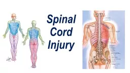
DEGENERATIVE SPINAL CORD DISEASES
Cemre
YILMAZ
Slide2Spinal Cord
The spinal cord extends from the foramen magnum where it continues with the medulla to the level of the first or second lumbar vertebra
.
It
terminates in a fibrous extension known as
filum
terminale
.
Terminal
portion of the spinal cord is called the
conus
medullaris
.
Spinal nerves pass through the vertebral column by exiting the intervertebral foramen. However, because the spinal cord does not reach the end of the vertebral column, the lumbar and sacral spinal nerves exit only by first going downward and traveling inferiorly through the vertebral canal before reaching their corresponding intervertebral foramina. For this reason, there is a collection of nerve roots at the lower end of the vertebral canal. This collection of nerve roots is called the
cauda
equina
due to a resemblance to a horse's tail
Slide3Slide4There are 31 pairs of spinal nerves
8 cervical
12
thoracal
5 lumbar
5 sacral
1 coccygeal
The spinal cord has two enlargements
Cervical(C3-T2)
:The cervical enlargement corresponds roughly to the brachial plexus nerves, which innervate the upper limb
Lumbar (T11-L1)
:The lumbar enlargement or lumbosacral enlargement corresponds to the lumbosacral plexus nerves, which innervate the lower limb
Slide5Spinal Cord
Slide6Vertebra
There are features that are common to all vertebral segments and others that are unique to each level. With the exception of C1, each segment has a vertebral body, which is the anterior portion of the vertebral segment. The superior and inferior portions of the vertebral body are referred to as the end plates which provide nutrition to the adjacent disk. The body is connected to the posterior elements by bilateral pedicles which are linear bony struts. The posterior elements consist of the pedicles, lamina, facets (articular process), transverse process and
spinous
process.
Slide7Slide8Slide9Slide10Intervertebral Discs
Each vertebral body segment(except C1-C2) is attached to the level above and below by an intervertebral disk
The disk has several functions:
1) It serves as a connection between the vertebral bodies
2) It acts as a pivot point
3) Distribute compressive forces
The disk is made of the nucleus
p
ulposus
and the annulus
fibrosus
Slide11Slide12Degenerative Spine Conditions
Herniated discs
Spinal stenosis
Degenerative disc
disease
Spondylo-lysis
/
listhesis
Degenerative scoliosis
Spondylosis
Slide13Risk Factors
a
ging
g
enetic
s
moking
weight
h
eavy lifting
s
edentary lifestyle
Slide14Symptoms
Degenerative spine conditions vary widely in their presentation. Some cause no symptoms at all.
When symptoms do occur, they often include
back pain or neck pain.
Other symptoms depend on the location and type of problem.
Slide15Disc Herniation
Disc herniation occurs when the annulus fibrous breaks open or cracks, allowing the nucleus
pulposus
to escape. This is called a herniated nucleus
pulposus
or herniated disc.
The most common sites are lumbar (L4-L5) herniated discs and cervical(C5-C6) herniated discs .Thoracic herniated discs are much less common.
Herniations
usually occur
posterlaterally
.
Slide16Slide17protrusion
:
base wider than herniation
confined to disc level outer
annular
fibres
intact
extrusion
:
base narrower than herniation
'
dome'may
extend above or bellow endplates or adjacent vertebrae
complete annular tear with passage of nuclear material beyond disc annulus
disc material can then migrate away from annulus or become sequestered
Sequestration
extruded disc material that has no continuity with the parent disc
is displaced away from the site of extrusion.
Slide18Cervical disc herniation
most common site C5-C6 / C6-C7
Pain (neck and upper extremities)
Numbness
Muscle weakness
Paresthesia
Urinary incontinence , loss of bowel control(rare)
Slide19Slide20Diagnosis
Physical exam
MRI
–
best
CT with
myelogram
–
more sensitive but invasive
X-ray
EMG
Treatment
Medication :NSAID
Physical therapy
Steroid injection
Surgery
Anterior
cervical discectomy and spine fusion (ACDF)
Posterior cervical discectomy
Cervical artificial disc replacement.
Slide21Lumbar Disc Herniation
Most common site L4-L5/L5-S1
Pain (lower back,
buttocks
,
lower
extremities)
Numbness
Foot drop
Cauda
equina
syndrome
Slide22Most commonly affected nerve sciatic nerve (L3-S1)
Slide23Straight
L
eg
R
aise
T
est
(
Lasegue’s
sign)
Neurologic pain which is reproduced in the leg and low back between 30-70 degrees of hip flexion is suggestive of lumbar disc herniation at the L4-S1 nerve roots.
Slide24Diagnosis
Physical
exam—straight leg raise test
MRI
CT
with
myelogram
X-ray
EMG
Treatment
Ice application
Medication : NSAID
,muscle relaxants
Heat therapy
Physical therapy
Steroid injection
Surgery
Microdiscectomy
Slide25Cauda Equina Syndrome(CES)
Cauda
equina
syndrome is caused by any narrowing of the spinal canal that compresses the
cauda
equina
nerve
roots
.
disc herniation
spinal
stenosis
traumatic
injury
tumors infectious conditions
arteriovenous
malformation or
hemorrhage
iatrogenic
injury
Slide26CES symptoms
Back pain
Saddle anesthesia
Sciatica pain
Bladder, bowel dysfunction
Gait disturbance
Anal and
achilles
reflex absent
Sexual dysfunction
Slide27Slide28Surgery indications
Severe pain
Progressive neurological deficit
Loss of bowel-bladder control
Slide29Slide30Spinal stenosis
Spinal stenosis is part of the aging
process
Progressive
narrowing of the spinal canal may occur alone or in combination with acute disc
herniations
. Congenital and acquired spinal
stenosis
place the patient at a greater risk for acute neurologic injury.
Spinal
stenosis is most common in the cervical and lumbar areas.
Slide31Spinal stenosis
Slide32Spinal stenosis
The most common reason to develop spinal stenosis is degenerative arthritis, or bony and soft tissue changes that result from
aging
.
The
normal "wear and tear" of
aging
can cause arthritis in the spine that leads to spinal stenosis. This can be from bone spurs (osteophytes) forming, bulging and wear of the intervertebral discs, and thickening of the ligaments between the vertebrae.
Slide33Spinal stenosis
Local and traveling pain, often described as a burning
sensation
Muscle weakness
Numbness
and
tingling
Loss
of fine motor
skills
Limited
mobility
Slide34Treatment
pain
medication
Exercise
Stretching
Hot
/cold
therapy
Epidural
steroid
injections
Lifestyle
changes like weight loss and quitting
smoking
Decompression surgery
Slide35Degenerative Disc Disease
Gradual
deterioration and thinning of the shock-absorbing intervertebral
discs by age
This condition can occur at any level of the spine
and
may cause a range of symptoms and intensity levels.
Unless
a degenerative disc places pressure upon an adjacent nerve, symptoms remain non-existent or strictly localized.
Slide36Degenerative Disc Disease
Pain
with activity
bending, lifting, and twisting
Severe
episodes of back or neck pain
(a
few days to a few months
Certain positions: sitting for
lumbar degenerative disc
pain
Slide37MRI Findings
Disc space narrowing
Fissures
, fluid, vacuum changes and calcification
Osteophytosis
Disk herniation
Malalignment
Stenosis
Slide38Slide39DDD Treatment
Pain control
Exercise and physical therapy
Lifestyle modifications
S
urgery
Slide40Spondylolysis
Caused by repeated
microtrauma
, resulting in
stress fracture of the pars
interarticularis
present
in ~5% of the population
%90 at the L5 level
higher
in the adolescent athletic
population
commonly asymptomatic
pain
with extension and/or rotation of the lumbar spine.
65% of patients with
spondylolysis
will progress to
spondylolisthesis
Slide41Spondylolysis
Plain
radiograph
oblique
limited
sensitivity compared to SPECT and CT
scotty
dog
sign
CT
/
MRI
Wide canal sign
Slide42Spondylolisthesis
is most frequent at L5/
S1
forward or backward slippage
the vertebra
Causes
of
spondylolisthesis
include trauma, degenerative,
tumor
and
birth defects
.
lower back or leg
pain, hamstring tightness, numbness
and tingling in the legs.
Slide43Treatment
Bracing to immobilize the spine for a short period
Pain
medications and/or anti-inflammatory
medication
Physical therapy
Decompressive
laminectomy
:reduces
irritation and inflammation in the area (but increases spinal instability
)
A
spinal fusion to provide stabilization of the affected area.
Slide44Spondylosis
Spinal osteoarthritis
With age, the bones and ligaments in the spine wear, leading to bone spurs
Over 80% of people over the age of 40 have evidence of
spondylosis
on X-ray
studies
Slide45Spondylosis
Neck/back pain
Stiffness
Paresthesia
weakness
Standing
Sitting
Sneezing
Coughing
Tilting neck backward
worsen the pain
Slide46Spurling’s
test(cervical compression test)
pain arising in the neck radiates in the direction of the corresponding dermatome
ipsilaterally
Shows cervical radiculopathy (many causes)
Lhermitte’s
sign
electric
shock-like sensation that occurs on flexion of the
neck
Reduced range of motion
MRI-CT
Slide47Treatment
(
NSAIDs
)
exercise
– such as swimming and
walking
Surgery
bowel
or bladder
dysfunction
spinal stenosis
neurologic dysfunctions
Unstable spine