

Agrawal Additional Professor Department of Ophthalmology AIIMS Rishikesh Acknowledgement Photographs in this presentation are courtesy of DrFreund K Bailey The Retinal Atlas2 ID: 932784
Download Presentation The PPT/PDF document "RETINAL DETACHMENT Dr.Ajai" is the property of its rightful owner. Permission is granted to download and print the materials on this web site for personal, non-commercial use only, and to display it on your personal computer provided you do not modify the materials and that you retain all copyright notices contained in the materials. By downloading content from our website, you accept the terms of this agreement.


Slide1
RETINAL DETACHMENT
Dr.Ajai
Agrawal
Additional
Professor
Department of Ophthalmology
A.I.I.M.S,
Rishikesh
Slide2Acknowledgement
Photographs in this presentation are courtesy
of
Dr.Freund
. K. Bailey (The Retinal Atlas,2
nd
Ed.)
and
Dr.Brad
Bowling (
Kanski’s
Clinical Ophthalmology,
8
th
Ed.)
Slide3Learning Objectives
At the end of the class, students shall be able to
Define and classify the various types of retinal detachments (R.D.)
Understand the pathophysiology and signs and symptoms of retinal detachments
Have a basic understanding of the management
of various types of retinal detachments
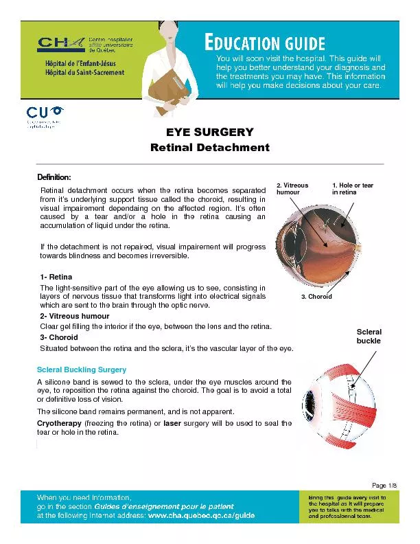
Slide4What is the retina?
Innermost coat of the eyeball.
Thin, delicate, transparent membrane.
Externally related to the choroid & sclera.
Thickness
Near optic disc – 0.56
mm
Equator – 0.18 to 0.2 mm
Ora-serrata
– 0.1 mm
Thinnest
at Fovea
.
Slide5Slide6Normal Fundus
Slide7Layers of the retina
Retinal Pigment Epithelium
Layer of Rods & Cones
External Limiting Membrane
Outer Nuclear Layer
Outer
Plexiform
Layer
Inner Nuclear Layer
Inner
Plexiform
Layer
Ganglion Cell Layer
Nerve
Fibre LayerInternal Limiting Membrane
Slide8RETINAL
DETACHMENT (RD
)
Definitions
and
classifications
Retinal breaks
Retinal detachment
Slide9Break - full-thickness defect in sensory retina
Hole - caused by chronic retinal atrophy
Tear - caused by dynamic vitreoretinal traction
d. Operculated
e. Dialysis
Morphology
of tears
a. Complete U-tear
b. Linear
c. Incomplete L-shaped
Definition and classification
Slide10DEFINITION
RETINAL DETACHMENT (
R.D.)
is defined as the
separation of neurosensory retina (NSR) from retinal pigment epithelium (RPE)
caused by breakdown of forces that attach the NSR to RPE resulting in
accumulation of sub retinal fluid (SRF) in the potential space
between the NSR and RPE.
Slide11Retinal detachment (RD)
Separation of sensory retina from RPE by subretinal fluid (SRF)
Rhegmatogenous
- caused by a
retinal break
Non-rhegmatogenous
- tractional or
exudative
Slide12Classification
Clinico
-etiologically
– Three types of retinal
detachment
Rhegmatogenous
(or
primary) retinal detachment
2.
Tractional retinal detachment3. Exudative retinal detachment
Slide13Classification-
Exudative
Tractional
Rhegmatogenous
Slide14Predisposing factors for RD
Myopia
Aphakia
(&
Pseudophakia
)
Trauma
Retinal Degenerations
PVD
Slide15Rhegmatogenous retinal detachment
Is usually associated with a retinal break
(hole/tear)
Sub
retinal
fluid(SRF) seeps and separates the neurosensory retina from the retinal pigment epithelium(RPE)
Slide16Indirect
ophthalmoscopy
Keep lens parallel to patient’s iris plane
Avoid tendency to move towards
patient
Ask the patient to move eyes and head
into optimal positions for examination
Technique
The higher the power, the less the
magnification, the shorter the working
distance
,
greater the field of view
Condensing lenses
Slide17Scleral indentation
Retinal breaks in detached
retina without indentation
Enhanced visualization of
breaks with indentation
Slide18Slitlamp
biomicroscopy
Goldmann
triple-mirror lens
Equatorial mirror (largest and
oblong
) - from 30 to equator
Peripheral mirror (square) -
from
equator to
ora
serrata
Gonioscopic
(smallest)
Image
is upside down
View of peripheral fundus
Slide19Predisposing peripheral degenerations
Slide20Typical lattice degeneration
Present in about 8% of general population
Present in about 40% of eyes with RD
Spindle-shaped islands of retinal thinning
Network of white lines within islands
Variable associated RPE changes
Small round holes within lesions are common
Overlying vitreous liquefaction
Exaggerated attachments
around margin of lesion
Retina
Vitreous
Slide21Snail track
degeneration
Indications for prophylaxis
- presence of holes
Sharply demarcated, frost-like bands
which are longer than lattice
Large round holes which carry
high risk of RD
Slide22White-without-pressure
Indications for prophylaxis
- giant tear in other eye
Translucent grey appearance of retina
Occasional giant tear formation along
posterior margin of lesion
Slide23Why is normal retina attached?
Vitreous
tamponade
Acid
mucopolysaccharides
(Bio glue)
Hydrostatic pressure( Less pressure in the sub retinal space)
RPE Pump
Slide24Pathogenesis
Retinal breaks are due to
dynamic
vitreoretinal traction
and
predisposing
retinal degenerationDegenerated fluid vitreous seeps through retinal break and collects as SRF between sensory retina and RPE leading to RD
Slide25Pathogenesis of
rhegmatogenous
RD
Possible sequelae of acute PVD
Two components for retinal break formation
Acute posterior vitreous detachment (PVD)
Predisposing peripheral retinal degeneration
Uncomplicated PVD (85%)
Retinal tear formation and
haemorrhage (10-15%)
Avulsion of retinal
vessel &
haemorrhage (uncommon)
Slide26Clinical features
Prodromal symptoms
Floaters (dark spots)
Photopsia
(flashes of light)
Symptoms of RD
Loss in the field of vision(Localised and relative progressing to total loss)
Painless loss of vision(usually rapid) with appearance of cloud/veil in front of affected eye
Slide27Slide28Signs of R.D.
External examination: Usually normal
Intra ocular pressure: Slightly lower or normal
Pupils: Normal reaction or
Relative Afferent Pupillary Defect
in extensive RD
Plane mirror examination: Greyish
reflex
Slide29Signs of R.D.
Ophthalmoscopy: Indirect Ophthalmoscopy with scleral indentation: Tobacco dust(Shafer’s sign)
Retinal breaks
Convex configuration with folds(corrugations)
Loss of the choroidal pattern
Retinal blood vessels - darker than in flat retina
Slide30Fresh
rhegmatogenous
RD - signs
Annual incidence - 1:10,000 of population
Eventually bilateral in 10%
Convex, deep mobile elevation
extending to ora serrata
Slightly opaque with dark blood vessels
Loss of choroidal pattern
Retinal breaks
Slide31Signs of old RD
Retinal thinning (due to atrophy)
Sub
retinal demarcation line/high water mark
(due to RPE proliferation)
Secondary intra retinal cysts
Slide32Longstanding
rhegmatogenous
RD - signs
Frequently inferior with small holes
Very thin retina
Secondary intraretinal cysts
Demarcation lines (high-water marks)
Slide33Investigations
Ultrasonography confirms the diagnosis
especially when media is hazy.
Visual field charting :
scotomas
(relative/absolute)
ERG: subnormal or absent
Slide34Complications
Proliferative
vitreo
retinopathy(PVR)
Complicated cataract
Uveitis
Phthisi
bulbi
Slide35Proliferative
vitreoretinopathy
Vitreous haze and
tobacco dust
Grade A (minimal)
Rigid retinal folds
Vitreous condensations
and strands
Grade C (severe)
Retinal wrinkling and
stiffness
Rolled edges of tears
Grade B (moderate)
Slide36Differential diagnosis of RD
Degenerative retinoschisis
Frequently bilateral
Smooth, thin and immobile
Occasionally breaks in one
or
both layers
Choroidal detachment
Associated with
hypotony
Unilateral, brown, smooth,
solid
and immobile
Ora
serrata
may be visible
Uveal effusion syndrome
Idiopathic
Rare, unilateral
Combined
choroidal
& exudative
detachments
Slide37Aims of management of RD
Seal/close retinal breaks
with
photocoagulation or
cryotherapy
(or diathermy – Jules
Gonin
-
Ignipuncture
)Sub Retinal Fluid
drainage
: for immediate apposition between sensory retina and RPE (Not in all cases)
Slide38Aims of management of RD
Maintain
chorioretinal
apposition/adhesion
by
Scleral Buckling to provide external tamponade
Pneumatic
retinopexy
Pars
plana
vitrectomy (to relieve traction on retina)
Slide39Technique of laser photocoagulation
Surround lesion with two rows of
confluent burns
Difficult for anterior lesions and if
media hazy
Slide40Technique of
cryotherapy
Surround lesion with single row of
cryo-applications
Preferred for treatment of large
areas
Slide41While viewing with indirect
ophthalmo
-
Scope indent
sclera gently
with
tip of
cryoprobe
Freeze break until sensory retina just
turns white
Cryotherapy
Slide42Drainage of
subretinal
fluid
Indications
Haemorrhage
Difficulty in localizing break
Immobile retina
Longstanding RD
Inferior RD
Retinal incarceration
Complications
Technique
Slide43Slide44Technique
(a) Cryotherapy
Pneumatic
retinopexy
Indications
RD with superior breaks
(b) Gas injection
(c) Postoperative positioning
(d) Flat retina
Slide45Vitrectomy
for giant tears
Unrolling of flap with light
pipe and probe
Completion of unrolling
Injection of silicone oil or
heavy liquid
Slide46Vitrectomy
for PVR
Dissection of star folds and peeling of
membranes
Injection of expanding gas or silicone oil
Slide47Tractional Retinal detachment
Occurs due to
mechanical
pull/traction on the retina by contraction of fibrous tissue in the vitreous.
Etiology
Proliferative Diabetic Retinopathy (PDR)
Penetrating posterior segment trauma
Retinopathy of prematurity
Slide48Signs of
tractional
RD
Slow progression and variable fibrosis
Does not extend to ora serrata
Concave, shallow immobile elevation
Highest at sites of
vitreoretinal
traction
Slide49Vitrectomy
for
tractional
RD
Release of circumferential
traction
Release of antero-
posterior traction
Endophotocoagulation
Slide50Exudative Retinal detachment
Occurs due to the retina being pushed away by a neoplasm or fluid accumulation beneath the retina following inflammatory or vascular lesions.
Slide51Pathogenesis and Causes of Exudative RD
Damage to RPE by
subretinal
disease
Passage
of fluid derived from choroid into
subretinal
space
1. Choroidal
tumours
Primary
Metastatic
2. Intraocular inflammation
Harada’s Disease
Posterior
Scleritis
3. Intraocular inflammation
Toxemia of pregnancy Hypoproteinemia
Slide52Pathogenesis and Causes of
Exudative
RD
4. Vascular
CSR
Coat’s disease
5. Iatrogenic
RD surgery
Excessive
retinal photocoagulation
6.
Miscellaneous
Choroidal neovascularization
Uveal effusion syndrome
Nanophthalmos
Slide53Signs of exudative RD
Convex, smooth elevation
May be very mobile and deep with
shifting fluid
Subretinal pigment (leopard spots)
after flattening
Slide54Medical Management
Inflammatory
conditions
(such
as
scleritis
and Vogt-
Koyanagi
-Harada
syndrome)
anti-inflammatory agents.Tumors- External
beam radiation therapy or brachytherapy with a plaque may be used for
choroidal
melanoma.
Metastatic
lesions respond to chemotherapy or localized radiation therapy.
Choroidal
hemangiomas may respond to laser photocoagulation or plaque brachytherapy. Retinoblastomas may be shrunk with chemotherapy and then treated locally with heat, laser, or cryotherapy.
Slide55Medical Management
Infectious
a
etiologies
-antibiotics.
Exudative retinal detachments secondary to chronic renal failure
may have
spontaneous retinal reattachment following renal transplant or renal dialysis.
Anti-VEGF agents -Coats disease.
Slide56Surgical Management
Conditions with vascular anomalies, such as Coats
disease-laser- cryotherapy
vitrectomy
Congenital
anomalies, such as optic pits or
colobomas
-
vitrectomy and
endolaser
techniques.
Slide57Differences between types of RD
Rhegmatogenous
Tractional
Exudative/serous
Hole/Break
+
-
-
Surface
Convex, corrugated
Concave, scalloped
Convex,
smooth
SRF shift
Rare
---
Shifting
SRF
Height of RD
Never reaches lens
Shallow
May reach/touch lens
Course
Progressive/Static
Progressive
Waxes/wanes
May
resolve by itself
Management
Surgical
Surgical
Medical/
s
urgical
Slide58Conclusion
Retinal Detachment is
defined as the separation of neurosensory retina (NSR) from retinal pigment epithelium (RPE
).
It may be
rhegmatogenous
,
tractional
or exudative.
Is one of the causes of significant visual loss.
Management is mainly surgical.