
3 acute myocardial infarction Ivan Čundrle Pavel Suk Jan Hruda ARK FNUSA 2016 Shock in general Shock Circulatory failure supply demand 1 cardiogenic pump 2 obstructive obstruction ID: 934404
Download Presentation The PPT/PDF document "1. hypovolemia/shock 2. pulmonary embol..." is the property of its rightful owner. Permission is granted to download and print the materials on this web site for personal, non-commercial use only, and to display it on your personal computer provided you do not modify the materials and that you retain all copyright notices contained in the materials. By downloading content from our website, you accept the terms of this agreement.


Slide1
1. hypovolemia/shock2. pulmonary embolism3. acute myocardial infarction
Ivan Čundrle, Pavel Suk, Jan Hruda
ARK, FNUSA
2016
Slide2Shock in general
Slide3ShockCirculatory failure – supply ≠ demand
1. cardiogenic – pump
2. obstructive – obstruction
3. hypovolemic – filling
4. distributive - shunts
1.
2.
4.
3.
Slide4Phases of Shock
Compensation
Decompensation
Refractor
y
Inflammatory cascade induction and organ damage - „secondary-hit model“
Organ damage further increases inflammatory cascade induction – vicious circle
Each type of shock differs at the beginning, however during the late phase all types of shock look similar (like distributive shock)
Slide5PathophysiologyThe main problem is cell
hypoxia
Stress response
catecholamines
, RAAS, cortisol, glucagon
Systemic inflammatory response
Imunity, inflamatory
mediatorsLocaly OK, but generalized response is harmful
Slide6Pathophysiology
1.
Macrocirculation
„blood flow centralization“
rarely „warm shock“
2. Microcirculation
Endothelium damage
Increased vascular leakage, leucocytes adherence
Main role in shock
3. Coagulation
Intravascular coagulation
4. Metabolism
Increased gluconeogenesis, proteolysis
Lactate acidosis
Slide7MODS
1.
Circulation
Vasoplegia
, cardiomyopathy
2. Lungs
ARDS
3. Kidney
AKI
4
.
Coagulation
DIC
5.
CNS
Altered consciousness
6.
GIT
Loss of barrier function
Slide8Signs/SymptomsNonspecific
Variable
Unreliable
Hypotension, tachycardia:
SBP < 90 mmHg
MAP < 60 mm Hg
Tf
> 100/min Cave compensatory shock/BB
Oliguria:
diuresis < 0,5 ml/kg/
hr
for 1 – 6hrs
Tachyp
nea
> 30
breaths/min
,
dyspnea
Skin:
Wet, cold
CRT(
>
2
s)
Mental state:
Confusion
Iritation
coma
Slide9Diagnostics1. Basic Lab
BC, coagulation
(Q/INR, aPTT, fib)
ions, gly
urea, kreatin
CRP
(sepsis?)
2.
ABGVentilation/oxygenation
Lac
, SvO2, ScvO2
Slide10ABGLac
Product of anaerobic glycolysis
Non-toxic, serves also as a fuel
normal
< 2
mmol/l Mortality predictorEarly sign
ScvO2
O2ER = (SaO2 – SvO
2) / SaO2, normaly 25%
normal
SvO
2
is 75
%
SvO
2
< 70
% =
O
2
supply impairment
Slide11Extended Hemodynamics
Slide12Initial resuscitation
Preload optimization
– increasing CO, fluids „volume challenge“, PLR
Persistent hypotension – catechol
s (
norepinephrine)If CO does not rise with fluids, add inotropes (dobutamin)
Lowering of inadequately high afterload (hypertension crisis)
Slide13Causal treatment
1. Cardiogenic shock
:
SCG - PCI
Arrhythmia treatment (AV block III., VT)
2. Hypovolemic shock: fluids
hemotherapy
damage control surgery/damage control resuscitation
3. Obstructive shock:
thrombolysis
Pericardial effusion evacuation
Slide141. Hypovolemic Shock
Slide15Most common Causes of Hypovolemia
B
leeding
Loss of fluids
(sweating, vomiting, diarrhea, ....) inadequate intake
Burns3rd space losses
Ileus
anafylaxis, sepsis (relativ hypovolemia)
Slide16Treatment
Initial resuscitation
Causal treatment
Goal is to restore organ perfusion, O2 supply
Early initiation
Secondary goal: restoration of O2 transportation capacity (ERY...)
Slide17Venous access2-3 thick peripheral cannulasCentral venous access is secondary (good for catecholamine, not fluids)
Exception: thick central lines (
Edwars
AVA 9F)
Slide18Arterial CatheterContinuous blood pressure monitoration
accurate
PPV
Repeated blood draws
Slide19SPV / PPV
Slide20SPV / PPV
Slide21Witch fluid to use?Ions Na
+
and K
+ - ICT/ECT distribution Oncotic pressure
plasma/ECT distribution
ICT 40%
ECT 20%
4%
Slide22GlucoseInadequate
Absolute water deficit
Hypernatremia correction
ICT 40%
ECT 20%
4%
Distribution volume
Slide23CrystaloidsFast leak into the ECT compartment
Substitution has to be 4x higher than the deficit (
...recently
questioned)
→ swellings
ICT 40%
ECT 20%
4%
Distribution volume
Slide24ColoidsDo not leave the intravascular compartmentEqual the deficit
Adverse reactions, contraindication – sepsis – renal damage
Good for acute blood loss
ICT 40%
ECT 20%
4%
Distribution volume
Slide25Blood productsOnly for blood loss corrections
5% albumin – natural colloid
expensive
Slide26Fluid resuscitation goals
Blood pressure, heart rate
Centralization reversal
diuresis
Decrease of the PPV / SVV
Sc(v)O2 a lactate normalizationFilling pressures(CVP, PAOP) not a good target
Slide27Acute bleedingBlood loss
15
%
(750 ml)
well compensated
30% (1,5 l) – tachycardia, oliguria, normotension – however ↓ organ perfussion
!More than 30%:
hypotension, tachycardia, oligo-anuria, ...
fractures
pelvis
(5000ml)
femur
(2000ml)
tibia
(1000 ml)
humerus
(800 ml)
radius
(400ml)
Slide28TreatmentBasic approach ... ABCD
Stop the bleeding
Give
i.v.
Fluids + catecholamineBlood type O- (4 immediately available), after 30 minutes type matched
Fresh frozen plasma 1:1 with erythrocytesTarget Hb 70-90 g/l, CNS trauma 100g/lThrombocytes 50 – 100 tis/ul
Fibrinogen 1,5 g/lPrevent hypothermia
, hypotension and acidosis
Slide292. Cardiogenic shock/ AIM
Slide30AIMMyocardial ischemia
Causes
Increased demand – tachycardia
Low oxygen content – anemia, CO poisoning, hypotension, pulmonary disease
Low coronary artery blood flow
90 % low coronary artery flow – coronary atherosclerosisTransmural ischemia – 3/4 of the myocardial wall (complete closure)Laminar/subendomyocardial
– 1/3 of the myocardial wall (partial closure + increased demand)
Slide31DiagnosticsPatinet history/clinical evaluation
ECG
a
Lab
ECHO, SCG
STEMI
NSTEMI
AP
History
Chest pain
Chest pain
Chest pain
ECG
ST elevation at least 2 mm in leads V1–V3 or at least 1 mm in V4–V6, I,
aVL
, II, III,
aVF
.
ST elevation in at least two adjacent leads.
New LBBB or (RBBB + LAH, RBBB + LPH).
ST depression at least 1 mm and /or T wave
inversion
ST depression at least 1 mm and /or T wave
inversion
Lab
Positive TNT
Positive TNT
Negative TNT
Slide32Localization
Anteroseptal
V1-V4
Anterolateral
V1-V6
Lateral
I,
aVL
, V5, V6
Lower/
diafragmatic
II, III,
aVF
Slide33Treatment
Continuous vital signs / ECG
IV access
Oxygen 4–8 l/min
12 lead ECG
Blood draw – Lab /TNT
Analgosedation
-
morphine
ASA 500 mg
i.v.
/200–400 mg
p.o.
heparin 5000 j
i.v.
/enoxaparin 1 mg/kg
s.c.
/
i.v.
clopidogrel
300
nebo
600 mg
p.o.
metoprolol
i.v.
If tachycardia
Slide34Cardiogenic ShockSevere, long-lasting arterial hypotensionLow CO
Increased filling pressure CVP/PAOP
Alteration of consciousness, oliguria, cold periphery, sweat, cyanosis
Slide35TreatmentMost important is to increase oxygen supply and lower oxygen consuption by myocardial muscle
Preload optimalization: diuretics/fluids
Afterload optimalization: vasodilatation / cave coronary arthery perfussion
Inotropy
– dobutamin
Treatment of the cause – PCI/thrombolysis
Slide36AvoidTachycardia – short diastolic phase, increased work load (however, sometimes only chance how to increase CO)
Severe hypotension, hypovolemia, vasodilatation
– low coronary artery perfusion pressure (
Ao
pressure – EDP LV)Increased preload/afterload
– increase of wall tension, work
Slide37TreatmentOxygen
– increase O2 supply
NIV,
invasive ventilation
– oxygenation, decreases preload/afterload
Diuretics/fluids – decrease preload, in later phase optimization of preload (fluid challenge/PLR)Catecholamine – norepinephrine for blood pressure, dobutamin (milrinon, levosimendan
) for inotropyVasodilatancia – nitrates, coronary artery, but also systemic vasculature ( increased blood pooling, preload lowering; arterial – afterload lowering)
Morphine – improves dyspnea
Slide383. Obstructive shock/ PE
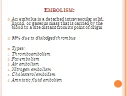
Slide39Pulmonary EmbolismSudden obstruction of pulmonary vasculature with emboli (blood cloth, fat, tumor, air/gas, foreign body, ...)
Etiology:
85% low extremity/pelvic DVT
Slide40Risc FactorsVirchov
trias
- venostasis, hypercoagulation
, vessel wall damageMajor surgeryLower extremity fracturesHypercoagulation (Leiden ...)Heart Failure (blood stasis)
Sepsis (coagulation activation)High age (70 years)Immobilization
ObesityPregnancyEconomy class syndromecorticoids, diuretics, HAC
Slide41DiagnosisHistory
Sudden dyspnea, chest pain, tachypnea, cough, syncope, hemoptysis
Clinical evaluation
tachypnea, cyanosis, hypotension, shock, tachycardia, neck veins distension
Lab
ABG - hypoxia, hypocapnea, RalcDD- negative – practically excludes PEDD- positive – tumors, inflammation, post-surgery, sepsis ...
Slide42EKG
Slide43Chest X-ray
Excludes other reasons for dyspnea
Fleischman sign- atelectasis
Westerman
sign – decreased pulmonary vascularization
Slide44ECHO
RV dilatation, paradoxical septum movements, pulmonary hypertension, Tri regurgitation
Slide45CT - AG
Slide46OtherVein US
–
femoral, popliteal
TEE
– thrombus in pulmonary arterySwan-Ganz - precapillary PH, high CVP, high RV pressure, increase PAP,
Ventilation/perfusion scan – low specificity
Slide47ManagmentClinical probability, DD, echo
and
CT
angio
Signs of DVT
3
Clinical probability:
low 0-1 (3,4%)
moderate 2-6 (20%)
high 7 (63%)
0-4 PE le
ss
probable
More than 4 - PE highly
probable
Other dg improbable
1,5
Tachycardia
100
1,5
Immobilization
more than
3 days, surgery within 4 weeks
1,5
DVT, PE in history
1,5
hemoptysis
1
malignancy
1
Slide481. High risk PE
(shock, hypotension)
CT angio
or ECHO, if CT unavailable/impermissible for the patient
CT/ECHO positive - trombolysis
Slide492. Low risk PE (without shock/hypotension)
High clinical suspicion – CT
angio
Low clinical suspicion – DD
Negative DD nearly completely exclude PE
TNT, NT pro BNP, RV dysfunction – thrombolysis/heparinization
Slide50Massive PE – unstable or RV dysfunction, TNT, NTproBNP
Thrombolysis
– optimally
within
48 hrs
alteplasis (0,9mg/kg)—10 mg bolus iv. + 90 mg cont. iv. for 2 hrs
+ heparin for min 72 hrs - UHF 80 IU/kg bolus + 18 IU/kg/
hr
Slide51Thrombolysis contraindications
Slide52Small PEUF heparin – bolus 80IU/kg + 18IU/kg/
hr
—
aPTT
1,5-2,5 times normAt least 6-10 days, than warfarinLMWH
- as effective as UHF, s.c. every 12 hrsAt least 6-10 days, than warfarinCave – renal dysfunction, antiXa (terap. 0,6-1,0 U/ml) 3
hrs after administration