
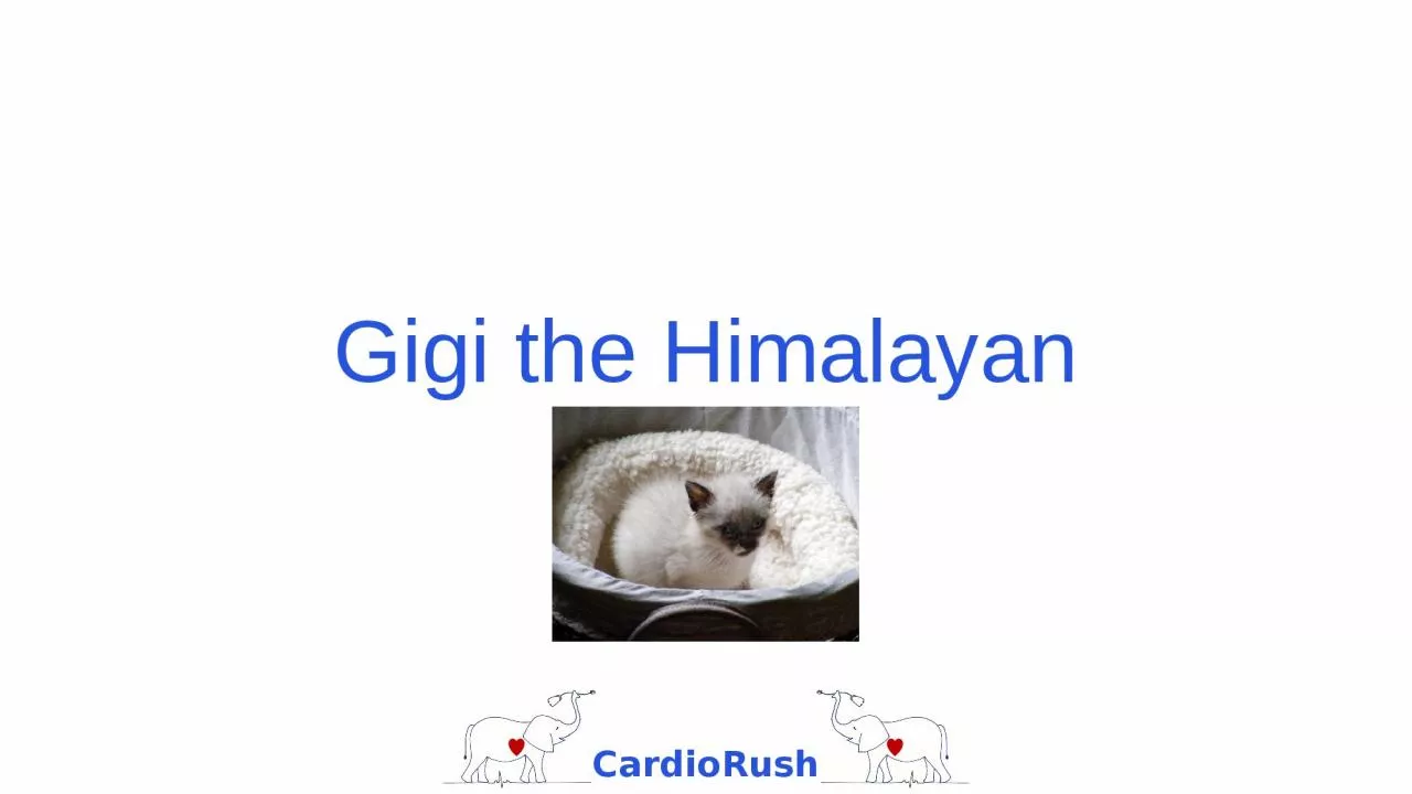
5 month old SF Himalayan History recent adoption no meds Presentation Gigi was presented for an overall health exam and a murmur was noted No clinical signs of heart disease were present other then mild shortness of breath after playing ID: 933381
Download Presentation The PPT/PDF document "Gigi the Himalayan Presentation" is the property of its rightful owner. Permission is granted to download and print the materials on this web site for personal, non-commercial use only, and to display it on your personal computer provided you do not modify the materials and that you retain all copyright notices contained in the materials. By downloading content from our website, you accept the terms of this agreement.


Slide1
Gigi the Himalayan
Slide2Presentation
5 month old SF Himalayan
History: recent adoption, no meds
Presentation: Gigi was presented for an overall health exam and a murmur was noted. No clinical signs of heart disease were present other then mild shortness of breath after playing.
Slide3Physical Exam
Heart
HR= 200 bpm
Murmur: 5/6 holosystolic murmur, PMI at the right sternal border
Gallop: loud S3 at left thorax
Arterial pulses-WNL; Jugular vein- distended 2/3 the way up the neckMucous membranes: pink with CRT under 2 secondsRespiratoryRR= 38/ minuteLung sounds were harsh, and Gigi was found to be tachypneic.
Slide4ECG at 25mm/sec
Slide5Thoracic Radiographs
Slide6Assessment
What is this patient’s heart rate and rhythm based on the ECG?
What other information can you gain from the ECG shown?
What is your radiographic interpretation?
Answers on next page
Slide7Assessment
What is this patient’s heart rate and rhythm based on the ECG?
220 bpm, sinus rhythm
What other information can you gain from the ECG shown?
There is a left ventricular enlargement pattern seen.
What is your radiographic interpretation?Generalized cardiomegaly – right and left heartPulmonary overcirculation with enlarged pulmonary vessels
Interstitial pattern compatible with
overcirculation
or mild pulmonary edema
Enlarged, rounded liver edges (relate this to jugular vein?)
Slide8Echocardiogram
Slide9Diagnosis
What type of heart disease does Gigi have?
Can you explain the radiographic pulmonary
overcirculation
and the enlarged pulmonary artery on echo?
Does Gigi have congestive heart failure?Answers on next page
Slide10Diagnosis
What type of heart disease does Gigi have?
Ventricular septal defect, high in the membranous portion of the IVS, below the aorta
Can you explain the radiographic pulmonary
overcirculation
and the enlarged pulmonary artery on echo?The VSD is left-to-right, so extra blood circulates to the main pulmonary artery, the pulmonary vasculature, the left atrium and left ventricle.Does Gigi have congestive heart failure?
The radiographs, hepatomegaly, S3 gallop, shortness of breath and jugular vein distension are all compatible with CHF.
Slide11What is your treatment plan?
Slide12Treatment Options
Medical and dietary management of congestive heart failure with furosemide, an angiotensin-converting enzyme inhibitor, maybe
pimobendan
, and dietary modification
Surgery – Pulmonary artery banding
Surgery – Cardiopulmonary bypass for patch repair of the VSD Surgery – Animals with VSD located lower in the muscular septum are candidates for a catheter-delivered device to occlude flow through the VSD – Gigi’s is too high in the IVS to allow this procedure