
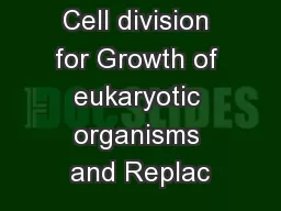
ANA 205 2 Differentiation development of a fertilized egg cell to form a complex multicellular organism involving cellular replication growth and progressive specialization for a variety of functions ID: 931263
Download Presentation The PPT/PDF document "Cell division- mitosis, meiosis , factor..." is the property of its rightful owner. Permission is granted to download and print the materials on this web site for personal, non-commercial use only, and to display it on your personal computer provided you do not modify the materials and that you retain all copyright notices contained in the materials. By downloading content from our website, you accept the terms of this agreement.


Slide1
Cell division- mitosis, meiosis , factors, specialization, functions, abnormal cell division, differentiation
ANA 205
Slide22
Differentiation
- development of a fertilized egg cell to form a complex, multicellular organism involving cellular replication, growth and progressive specialization for a variety of functions.
The fertilized egg (
zygote
) divides by
mitosis
to produce two genetically identical daughter cells, each of which divides to produce two more daughter cells and so on.
These daughter cells specialize and produce the
terminally differentiated
cells of mature tissues, such as muscle or skin cells.
Slide33
Most tissues retain a population of undifferentiated cells (
stem cells
) that are able to divide and replace the differentiated cell population as required.
The interval between mitotic divisions is known as the
cell cycle
.
All body cells divide by
mitosis
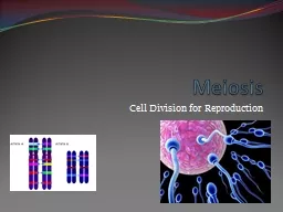
except for male and female germ cells, which divide by
meiosis
to produce
gametes
Slide44
Adult cell population
Terminally differentiated cells of some tissues, such as the
neurons
, lose the ability to undergo mitosis.
Stem cells of gut and skin
undergo continuous cycles of mitotic division throughout the lifespan of the organism replacing cells lost during normal wear and tear.
Between these extremes are cells such as
liver cells
that do not normally divide but retain the capacity to undergo mitosis should the need arise –
Facultative dividers
.
Cell division and differentiation are balanced by
cell death
in all organisms. In these circumstances, cell death occurs by a mechanism known as
apoptosis.
Slide55
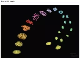
Cell division, or Mitosis
Can be observed with the light microscope.
Longitudinal duplication of the chromosomes occurs
Chromosomes are distributed to the daughter cells.
Phase between two mitoses is
interphase
.
Division of
somatic cells
(all body cells except for the germ cells) occurs in two phases.
a. chromosomes duplicated in S phase are distributed equally between the two potential daughter cells identical to that of the parent cell
b. dividing cell is cleaved into genetically identical daughter cells by
cytoplasmic
division or
cytokinesis
.
Always equal and symmetrical,
cytokinesis
may result in the formation of two daughter cells with grossly unequal amounts of cytoplasm or
cytoplasmic
organelles.
- May occur in the absence of
cytokinesis
resulting in the formation of
binucleate
and multinucleate cells.
A continuous process divided into four phases,
prophase
,
metaphase
,
anaphase
and
telophase
.
Slide66
Interphase
Duplication of the
centrosomes
and
centrioles
starts in the
interphase
, before mitosis.
Centrosome
divides during
interphase
,
Divided into three phases: G
1
(
presynthesis
), S (DNA synthesis), and G
2
(post-DNA duplication).
S phase is characterized by the synthesis of DNA and the beginning of the duplications of the
centrosomes
with their
centrioles
.
Slide77
Prophase
Chromosomes become visible within the nucleus.
Gradual coiling of nuclear chromatin
Chromosomes are condensed and shortened
Centrosomes
with their
centrioles
separate,
and
migrate to each pole of the cell. Microtubules of the mitotic spindle appear between the two centrosomes (interpolar microtubules)Nucleolus disintegrates.Cell division requires the presence of a structure called the mitotic apparatus - a spindle of longitudinally arranged microtubules between a pair of centrioles at each pole of the dividing cell. Microfilaments and microtubules of the cytoskeleton disaggregate into their protein subunits. At the end of prophase, the nuclear envelope is broken by phosphorylation of the nuclear lamina proteins.
Slide88
Metaphase
Nuclear envelope disintegrates
Chromosomes migrate to the equatorial plane of the cell
At the poles, each chromosome divides longitudinally to form two chromosomes called sister
chromatids
.
Chromatids
attach to the microtubules of the mitotic spindle at the
kinetochore
, located close to the
centromere
.
Mitotic spindle moves into the nuclear area and each duplicated chromosome becomes attached at the kinetochore. Kinetochore is a DNA and protein structure on each duplicated chromosome located at the centromere [binds the duplicated chromosomes (chromatids) together]. Controls entry of the cell into anaphase so that the process of mitosis does not progress until all chromatid pairs are aligned at the cell equator - metaphase checkpoint Prevents the formation of daughter cells with unequal numbers of chromosomes. Chromosomes become arranged in the plane of the spindle equator known as the
equatorial
or
metaphase plate.
Slide99
Anaphase
Sister
chromatids
separate
(split at the
centromere
) from
each
other
Migrate
toward the opposite poles of
the
cell, pulled by microtubules. Centromeres move away from the center, pulling the remainder of the chromosome along. Centromere is the constricted region of a mitotic chromosome that holds the two sister chromatids together.Mitotic spindle becomes lengthened by addition of tubulin subunits to its interpolar microtubules while astral microtubules joining the centrosome to the cell cortex shorten.
Centrioles
are pulled apart and the
chromatids of each duplicated chromosome are drawn to opposite ends of the spindle.
By the end of anaphase, two groups of identical chromosomes are clustered at opposite poles of the cell.
Slide1010
Telophase
Characterized by the reappearance of nuclei in the daughter cells.
Chromosomes revert to their
semidispersed
state, nuclear envelope reassembles and nucleoli again become apparent.
Chromosomes begin to uncoil and to regain their
interphase
conformation.
Plane of
cytoplasmic
division is defined by the spindle equator, thus producing two cells of equal size.
Plasma membrane around the spindle equator becomes indented to form a circumferential furrow - the cleavage furrow, which is cleaved into two daughter cells. Cytokinesis occurs as a result of contraction of a ring of microfilaments present beneath the surface of the cleavage furrow. In early G1 phase, the mitotic spindle disaggregates and centrioles duplicate in preparation for the next mitotic division.
Slide1111
Turnover rate of cells
Most tissues undergo constant cell turnover because of continuous cell division and the ongoing death of cells.
Nerve tissue and cardiac muscle cells are exceptions, since they do not multiply
postnatally
and therefore cannot regenerate.
The turnover rate of cells varies greatly from one tissue to another (rapid in the epithelium of the digestive tract and the epidermis and slow in the pancreas and the thyroid gland).
Slide1212
Cell
Cycle
DNA replication occurs
during
interphase
,
when no visible phenomena of cell division can be seen with the microscope.
Alternation
between mitosis and
interphase
, known as the
cell cycle,
occurs in all tissues with cell turnover. Cell cycle can be divided into two stages: mitosis, consisting of the four phases: (prophase, metaphase, anaphase, and telophase), and interphase (Go, S, G1, G2).
Slide1313
The
four phases of the cell cycle
.
G
1
phase (
presynthesis
)
Varies in duration, in bone tissue, G
1
lasts 25 h. The S phase (DNA synthesis) lasts about 8 h. The G
2
-plus-mitosis phase lasts 2.5-3 h.Depends on the rate of cell division in the tissue.Cell either continues the cycle or enters a quiescent phase called G0. The checking or restriction point (R) in G1 stops the cycle under conditions unfavorable to the cell. When the cell passes this restriction point, it continues the cycle through the synthetic phase (S) and the G2 phase, originating two daughter cells in mitosis except when interrupted by another restriction point.
Slide1414
Intense synthesis of RNA and proteins, previously reduced to one-half by mitosis, is restored to its normal size.
In cells that are not continuously dividing, the activities of the cell cycle may be temporarily or permanently suspended.
Go Phase
Cell either continues the cycle or enters a
quiescent phase called G
0
.
Cells in such a state (
eg
, muscle, nerve) are referred to as being in the G
0
phase.
Slide1515
G2 phase
Accumulation of energy to be used during mitosis
Synthesis of
tubulin
to be assembled in mitotic microtubules
Synthesis of chromosomal
nonhistone
proteins.
A checkpoint at which the cell remains until all DNA synthesized with defects is corrected.
Accumulation of the protein complex maturation promoting factor (
MPF
) that induces the beginning of mitosis, the condensation of the chromosomes, the rupture of the nuclear envelope, and other events related to mitosis.
Slide1616
A dividing phase (
M phase
) and a non-dividing phase (
interphase
), which usually occupies most of the life cycle of the cell.
A discrete period during
interphase
when nuclear DNA is replicated is the synthesis or
S phase
, is completed some time before the onset of mitosis.
Interphase
may be divided into three separate phases. Between the end of the M phase and the beginning of the S phase is the first gap or G1 phase; usually longer than the other phases of the cell cycle.
During the G
1
phase, cells differentiate and perform their specialized functions as part of the whole tissue.
The interval between the end of the S phase and the beginning of the M phase, the second gap or
G
2
phase
, is short and is the period in which cells prepare for mitotic division.
Cell Cycle
Slide1717
MEDICAL APPLICATION
Proteins that stimulate mitotic activity:
nerve growth factor, epithelial growth factor, fibroblast growth factor
and
erythropoietin
.
Cell
cycle
is
regulated by
DNA
damage
which arrests the cell cycle not only in G2 but also at a checkpoint in G1.In mammalian cells, arrest at the G1 checkpoint is mediated by the action of a protein known as p53. Inheritance of damaged DNA by daughter cells results in mutations and instability of the genome leading to the development of cancer.Rapidly growing tissues frequently contain cells in mitosis, whereas slowly growing tissues do not.
Slide1818
Normal cell proliferation and differentiation are controlled by a group of genes called
protooncogenes
Cell proliferation is regulated by mechanisms that stimulate or retard mitosis according to the needs of the organism.
Chemical substances, radiation and viral infections can induce DNA damage, mutation, and abnormal cell proliferation result in the formation of tumors.
Tumor
- any localized swelling in the body caused by inflammation or abnormal cell proliferation.
Neoplasm
- abnormal mass of tissue formed by uncoordinated cell proliferation; either benign - slow growth and no invasiveness or malignant - rapid growth and great capacity to invade tissues and organs.
Cancer
is the common term for all malignant tumors
Slide1919
Chromosomes during mitosis
The nuclei of all somatic cells of an individual contain
deoxyribonucleic acid (DNA)
, called the
genome
.
The DNA is arranged into chromosomes consisting of
deoxyribonucleotides
with a double-stranded
(helical) structure
.
Each strand consists of alternating
deoxyribose S and phosphate P moieties. Each deoxyribose unit is covalently bound to a purine or pyrimidine base, which is in turn non-covalently linked to a complementary base on the other strand, thus linking the strands together. The bases are of four types, adenine A, cytosine C, thymine T and guanine G,
Slide2020
Slide2121
46
chromosomes (the
diploid
number) comprising 22 homologous pairs, the
autosomes
, and 2
sex chromosomes
, either XX in the female or XY in the male.
During S phase, each chromosome is duplicated.
Identical chromosomes known
as
chromatids are attached to one another at the centromereDNA molecule in each chromosome binds to histone proteins and hold the chromosome within the nucleus. Karyotyping - examination of the chromosomes of dividing cells- Gives diagnostic information about the chromosomal complement of an individual or of a malignant tumour
Slide2222
Karyotype
Slide2323
Stem cells
- dividing cells / undifferentiated cells in tissues with a regular turnover of cells,
Totipotent
- embryonic stem cells that able to differentiate into any other cell type
Multipotent
- stem cells found in adults that are able to produce cells of several lineages
eg
haemopoeitic
stem cell
Unipotent - producing only a single cell type eg epidermal stem cells of the skin that produce only epithelial cells.The advantages of such cells are in the treatment of degenerative diseases - being able to grow a new kidney or even a new limb to order. - Ethical minefield, especially the use of embryonic cells.
Slide2424
Comparison of mitosis and meiosis
The progress of only one homologous pair is represented here.
The key differences between the two forms of cell division are as follows: Meiosis involves one reduplication of the chromosomes followed by two sequential cell divisions.
Thus a diploid cell produces four haploid germ cells (gametes).
Crossing over occurs only in meiosis, to rearrange alleles such that every gamete is genetically different. In contrast, the products of mitosis are genetically identical.
Slide2525
Abnormal cell division
Meiotic cell division sometimes malfunction.
Trisomies
Failure of the homologous chromosome pairs to separate during the first meiotic division (
nondisjunction
) or failure of the two sister
chromatids
to separate during the second meiotic division (
anaphase lag
).
The resulting gamete has two copies of the relevant chromosome and when
fertilisation occurs a third copy is added usually from the motherKaryotyping detects cytogenetic abnormalities by the techniques of amniocentesis or chorionic villous sampling.Trisomy 21 (three copies of chromosome 21, also known as Down syndrome); increased incidence with increasing maternal age. Trisomy
18 (Edward syndrome)
Trisomy
13 (Patau syndrome) give rise to live births.
Slide2626
Apoptosis
Essential
part of normal fetal development, growth of juveniles and control of cell numbers in adults, where it balances cell division.
Apoptosis also occurs in a number of pathological conditions.
Examples
: epithelial cells in the skin or the lining of the gastrointestinal tract, developing T lymphocytes that are capable of reacting to normal body components are triggered to self-destruct in the thymus, in humans the webs between the fingers and toes disappear and the tadpole loses its tail as it matures into a frog, growth of ovarian follicles before ovulation in females followed by regression of the corpus
luteum
by apoptosis to form a corpus
albicans
.
Failure of apoptosis may be as important in cancer as unrestricted cell division.
Slide27MEDICAL APPLICATION
Prevents
the proliferation of malignant cells that develop as a result of accumulated mutations in the DNA
.
First
discovered in developing embryos, where programmed cell death is an essential process for shaping the embryo (
morphogenesis); also
a common event in the tissues of normal adults.
Necrosis
: the
accidental death of cells, a pathological
process
.
27