
WS 201920 lecture 16 1 Bioinformatics III What are microRNAs How can one identify microRNAs What is the function of microRNAs Laird Hum Mol Gen 14 R65 2005 Huntzinger Izaurralde Nat Rev Genet 12 99 2011 ID: 933299
Download Presentation The PPT/PDF document "V16: involvement of microRNAs in GRNs" is the property of its rightful owner. Permission is granted to download and print the materials on this web site for personal, non-commercial use only, and to display it on your personal computer provided you do not modify the materials and that you retain all copyright notices contained in the materials. By downloading content from our website, you accept the terms of this agreement.


Slide1
V16: involvement of microRNAs in GRNs
WS 2019/20 - lecture 16
1
Bioinformatics III
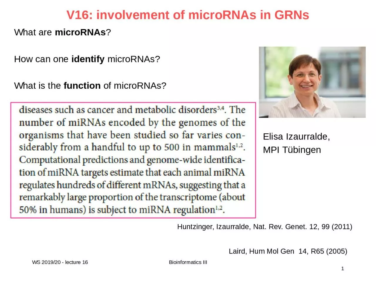
What are microRNAs?How can one identify microRNAs?What is the function of microRNAs?
Laird, Hum Mol Gen 14, R65 (2005)
Huntzinger, Izaurralde, Nat. Rev. Genet. 12, 99 (2011)
Elisa Izaurralde,
MPI Tübingen
Slide22
RNA world
short name full name function oligomerization
mRNA, rRNA, tRNA, you know them well ... Single-strandedsnRNA small nuclear RNA splicing and other functionssnoRNA small nucleolar RNA nucleotide modification of RNAsLong ncRNA Long noncoding RNA variousmiRNA microRNA gene regulation single-strandedsiRNA small interfering RNA gene regulation double-stranded
WS 2019/20 - lecture 16
2
Bioinformatics III
Slide33
RNA double-strand structure
P
NAS (2014) 111, 15408–15413.
RNA, like DNA, can form double helices held together by the pairing of complementary bases, and such helices are ubiquitous in functional RNAs.In contrast to DNA, RNA forms an A-form helix with a radius of ∼1.2 nm and a length increase per base pair of ∼2.8 Å, ∼20% wider and shorter than B-form dsDNA
WS 2019/20 - lecture 16
3
Bioinformatics III
Slide44
RNA
secondary structure
Basic structural motifs of RNA secondary structure. This RNA consists of five stems (labeled S1-S5) connected by loops (labeled according to loop type).
WS 2019/20 - lecture 16
4
Bioinformatics III
Slide55
Structure of single-stranded RNA
www.rcsb.org
Also single stranded RNA molecules frequently adopt a specific
tertiary structure. The scaffold for this structure is provided by secondary structural elements which are non-covalent hydrogen bonds within the molecule. This leads to several recognizable structural "domain“ types of secondary structure such as hairpin loops, bulges and internal loops.RNA hairpin 2RLU Stem loop 1NZ1
WS 2019/20 - lecture 16
5
Bioinformatics III
Slide66
RNA
tertiary structure
Suslov et al. Nature Chemical Biology 11, 840–846 (2015).
3D structure of the VS ribozyme. This ribozyme (ribonucleic acid & enzyme) from the mitochondria of Neurospora performs self-cleavage during replication. Shown is the catalytic domain
(helices 2–6) of one protomer and the substrate-helix (helix 1) that belongs to another protomer. The three-way helical junctions 2-3-6 and 3-4-5 organize the overall fold of the catalytic domain.
WS 2019/20 - lecture 16
6
Bioinformatics III
Yellow
spheres
:
scissile
phosphate
.
Red
sticks
:
catalytic
nucleobases
.
Junction
1-2-7
and
accompanying
helices
1
and
7
have
been
omitted
for
clarity
.
Slide77
snRNAs
www.wikipedia.org
Small nuclear RNA (
snRNA) are found within the nucleus of eukaryotic cells. They are transcribed by RNA polymerase II or RNA polymerase III and are involved in a variety of important processes such as RNA splicing, regulation of transcription factors or RNA polymerase II, and maintaining the telomeres. snRNAs are always associated with specific proteins. The snRNA:protein complexes are referred to as small nuclear ribonucleoproteins (snRNP) or sometimes as snurps.5 small nuclear RNAs (snRNAs) and approximately50 different proteins make up the splicing machinery. The five snRNAs are essential splicing factors.
Each snRNA is associated with several different proteins to make up five snRNP complexes, called U1, U2, U4, U5 and U6.
WS 2019/20 - lecture 16
7
Bioinformatics III
Slide88
snoRNAs
www.wikipedia.org
A large
subgroup of snRNAs are known as small nucleolar RNAs (snoRNAs). These are small RNA molecules that play an essential role in RNA biogenesis and guide chemical modifications of rRNAs, tRNAs and snRNAs. They are located in the nucleolus and the cajal bodies of eukaryotic cells.Predicted structure of hybrids between novel snoRNAs and target RNAs. Top: predicted snoRNA Bottom: target small nuclear RNA (snRNA)Kishore et al. Genome Biology 2013 14:R45
WS 2019/20 - lecture 16
8
Bioinformatics III
Slide99
RNA interference
www.wikipedia.org
RNA interference may involve siRNAs or miRNAs.
Nobel prize in Physiology or Medicine 2006 for their discovery of RNAi in C. elegans in 1998. Andrew Fire Craig Mello
WS 2019/20 - lecture 16
9
Bioinformatics III
Slide1010
siRNAs
www.wikipedia.org
Small interfering RNA (
siRNA), sometimes known as short interfering RNA or silencing RNA, is a class of double-stranded RNA molecules, that are 20-25 nucleotides in length (often precisely 21 nt) and play a variety of roles in biology. Most notably, siRNA is involved in the RNA interference (RNAi) pathway, where it interferes with the expression of a specific gene. In addition to their role in the RNAi pathway, siRNAs also act in RNAi-related pathways, e.g., as an antiviral mechanism or in shaping the chromatin structure of a genome.
WS 2019/20 - lecture 16
10
Bioinformatics III
Slide1111
miRNAs
www.wikipedia.org
In contrast to double-stranded siRNA,
microRNAs (miRNA) are single-stranded RNA molecules of 21-23 nucleotides in length.miRNAs have a crucial role in regulating gene expression. Remember: miRNAs are encoded by DNA but not translated into protein (non-coding RNA).
WS 2019/20 - lecture 16
11
Bioinformatics III
Slide12Bioinformatics III
Overview of the miRNA network
Ryan et al. Nature Rev. Cancer (2010) 10, 389
WS 2019/20 - lecture 16
12
RNA polymerase II (Pol II) produces a 500–3,000 nucleotide transcript, called the primary microRNA
(pri-miRNA).
AA, poly A tail;
m7G, 7-methylguanosine cap; ORF, open reading frame.
pri-miRNA is then cropped to form a pre-miRNA hairpin of ~60–100 nucleotides in length by a multi-protein complex that includes the protein DROSHA.
Slide13Bioinformatics III
Overview of the miRNA network
Ryan et al. Nature Rev. Cancer (2010) 10, 389
WS 2019/20 - lecture 16
13
This double-stranded
pre-miRNA hairpin structure is exported from the nucleus by RAN GTPase and exportin 5 (XPO5). Finally, the pre-miRNA is cleaved by the protein DICER1 to produce two miRNA strands:- a mature miRNA sequence, approximately 20 nt in length, - and its short-lived complementary sequence, which is denoted miR.
Slide14Bioinformatics III
DROSHA X-ray structure
WS 2019/20 - lecture 16
14
AA, poly A tail;
m7G, 7-methylguanosine cap; ORF, open reading frame.
The overall structure of DROSHA is surprisingly similar to that of Dicer despite no sequence homology apart from the C-terminal part.
This suggests that DROSHA may have evolved from a Dicer homolog.
Kwon et al. Cell. (2016) 164:81-90.
Slide15Bioinformatics III
Overview of the miRNA network
Ryan et al. Nature Rev. Cancer (2010) 10, 389
WS 2019/20 - lecture 16
15
The RISC complex is then targeted by the miRNA to the target 3′ untranslated region of a mRNA sequence to facilitate
repression and cleavage. The main function of miRNAs is to down-regulate gene expression of their target mRNAs.
The thermodynamic stability of the miRNA duplex termini and the identity of the nucleotides in the 3′ overhang determines which of the single strand miRNA is incorporated into the RNA-inducing silencing complex (
RISC).
Slide1616
miRNAs
www.wikipedia.org
Mature miRNA molecules are partially complementary to
one or more mRNA molecules. Fig. shows the solution NMR-structure of let-7 miRNA:lin-41 mRNA complex from C. elegans Cevec et al. Nucl. Acids Res. (2008) 36: 2330. miRNAs typically have incomplete base pairing to a target and inhibit the translation of many different mRNAs with similar sequences. In contrast, siRNAs typically base-pair perfectly and induce mRNA cleavage only in a single, specific target.
WS 2019/20 - lecture 16
16
Bioinformatics III
Slide1717
discovery of let7
WS 2019/20 - lecture 16
17
Bioinformatics III
Pasquinelli et al. Nature (2000) 408, 86
www.wikipedia.org
The first two known microRNAs, lin-4
and let-7, were originally discovered in the nematode C. elegans. There, they control the timing of stem-cell division and differentiation. let-7 was subsequently found as the first known human miRNA. let-7 and its family members are
highly conserved across species in sequence and function. Misregulation of let-7 leads to a less differentiated cellular state and the development of cell-based diseases such as cancer.
Slide1818
miRNA discovery
miRNA discovery approaches, both biological and bioinformatics,
have now yielded many thousands of miRNAs. This process continues with new miRNA appearing daily in various databases.miRNA sequences and annotations are compiled in the online repository miRBase (http://www.mirbase.org/). Each entry in the database represents a predicted hairpin portion of a miRNA transcript with information on the location and sequence of the mature miRNA sequence
WS 2019/20 - lecture 16
18
Bioinformatics III
Liu et al. Brief Bioinf. (2012) doi: 10.1093/bib/bbs075
Slide1919
miRNAs recognize targets by Watson-Crick base pairing
(a)
Plant miRNAs recognize fully or nearly complementary binding sites.(b) Animal miRNAs recognize partially complementary binding sites which are generally located in 3’ UTRs of mRNA.Complementarity to the 5’ end of the miRNA – the “seed” sequence containing nucleotides 2-7 – is a major determinant in target recognition and is sufficient to trigger silencing.
WS 2019/20 - lecture 16
19
Bioinformatics III
Huntzinger, Izaurralde, Nat. Rev. Genet.
12, 99 (2011)
4
6 = (22)6 = 212 = 4096 k-mers of length 6On average, the 3'-UTR in humans is ca. 800 nt long (www.wikipedia.org)20.000 genes x 800 nt / 4096 6-mers = 4000 binding sites for 1 miRNA 6-mer
Slide2020
Bioinformatics prediction of miRNAs
With bioinformatics methods, putative miRNAs are first predicted
in genome sequences based on the structural features of miRNA. These algorithms essentially identify hairpin structures in non-coding and non-repetitive regions of the genome that are characteristic of miRNA precursor sequences. The candidate miRNAs are then filtered by their evolutionary conservation in different species. Known miRNA precursors play important roles in searching algorithms because structures of known miRNA are used to train the learning processes to discriminate between true predictions and false positives.Many algorithms exist such as miRScan, miRSeeker, miRank, miRDeep, miRDeep2 and miRanalyzer.
WS 2019/20 - lecture 16
20
Bioinformatics III
Liu et al. Brief Bioinf. (2012) doi: 10.1093/bib/bbs075
Slide2121
Recognition of miRNA targets
There seem to be two classes of binding patterns.
One class of miRNA target sites has perfect Watson–Crick complementarity to the 5’-end of the miRNAs, referred to as ‘seed region’, which includes positions 2–7 of miRNAs.When bound in this way, miRNAs suppress their targets without requiring significant further base pairings at the 3’-end of the miRNAs.
WS 2019/20 - lecture 16
21
Bioinformatics III
Liu et al. Brief Bioinf. (2012) doi: 10.1093/bib/bbs075
The second class of target sites has imperfect complementary base pairing at the 5’-end of the miRNAs, but it is compensated via additional base pairings in the 3’-end of the miRNAs.
The multiple-to-multiple relations between miRNAs and mRNAs lead to complex miRNA regulatory mechanisms.
Slide22Bioinformatics III
miRNA-target prediction algorithms
Liu et al. Brief Bioinf. (2012) doi: 10.1093/bib/bbs075
WS 2019/20 - lecture 16
22
Slide23Bioinformatics III
Predicting miRNA function based on target genes
Liu et al. Brief Bioinf. (2012) doi: 10.1093/bib/bbs075
WS 2019/20 - lecture 16
23
The most straight-forward approach for miRNA functional annotation is through
functionalenrichment analysis using the miRNA-target genes.
This approach assumes that miRNAs have similar functions as their target genes.
Slide24Bioinformatics III
Predicting miRNA function based on correlated expression
Liu et al. Brief Bioinf. (2012) doi: 10.1093/bib/bbs075
WS 2019/20 - lecture 16
24
miRNA functional annotation heavily relies on the miRNA-target prediction.
In the last few years, many studies have been conducted to infer the miRNA regulatory mechanisms by incorporatingtarget prediction with other genomics data, such as
the expression profiles of miRNAs and mRNAs.
Slide25Bioinformatics III
Discovering MRMs
Liu et al. Brief Bioinf. (2012) doi: 10.1093/bib/bbs075
WS 2019/20 - lecture 16
25
A MRM (
group of co-expressed miRNAs and mRNAs) may be defined as a special bipartite graph, named biclique, where two sets of nodes are connected by edges. Every node of the first set representing miRNA is connected to every node of the second set representing mRNAs. The weights of edges correspond to the miRNA–mRNA binding strength are
inferred from target prediction algorithmsMost of the integrative methods for MRM discovery are based on the assumption that miRNAs negatively regulate their target mRNAs so that the expression of a specific miRNA and its targets should be anti-correlated.
Slide26Bioinformatics III
miRNA-mRNA network
Liu et al. Brief Bioinf. (2012) doi: 10.1093/bib/bbs075
WS 2019/20 - lecture 16
26
Up-regulated miRNAs are coloured in
red and down-regulated miRNAs are coloured in green. Up-regulated mRNAs are coloured in
yellow, while down-regulated mRNAs are coloured in blue.
A MRM identified from analysis of schizophrenia patients. It shows that miRNAs may up/down regulate their target mRNAs, either directly or indirectly.
Slide27Bioinformatics III
Volinia et al. PNAS (2013) 110, 7413
WS 2019/20 - lecture 16
27
FFL:
feed
-forward
loop FBL: feedback loop
Slide28Bioinformatics III
WS 2019/20 - lecture 16
28
Slide29Bioinformatics III
WS 2019/20 - lecture 16
29
Slide30Bioinformatics III
WS 2019/20 - lecture 16
30
Slide31Bioinformatics III
WS 2019/20 - lecture 16
31
Slide32Bioinformatics III
WS 2019/20 - lecture 16
32
Slide3333
TFmiR
Hamed et al.
Nucl Ac Res
43: W283-W288 (2015)
WS 2019/20 - lecture 16
Bioinformatics III
Slide3434
TFmiR
Hamed et al.
Nucl Ac Res
43: W283-W288 (2015)
WS 2019/20 - lecture 16
Bioinformatics III
Slide35Bioinformatics III
Significance of FFL motifs
WS 2019/20 - lecture 16
35
Compare how often FFL motifs appear in the real network to the number of times they appear in randomized ensembles preserving the same node degrees.
Use degree preserving randomization algorithm.For 2 × L steps, two edges e1 = (v1, v2) and e2 = (v3, v4) are randomly chosen from the network and rewired such that the start and end nodes are swapped, i.e. e3 = (v1, v4) and e4 = (v3, v2) if {
e3, e4} ∈ V.Construct 100 random networks. Compare motif frequencies to the real network. The P-value is calculated aswhere Nh is the number of random times that a certain motif type is acquired more than or equal to its number in the real network, and Nr is 100.
Slide36Bioinformatics III
Enriched motifs
WS 2019/20 - lecture 16
36
We identified 53 significantly enriched FFL motifs in breast cancer GRN:
-3 compositeFFLs, - 2 TF-FFLs, - 6 miRNA-FFLs - 42 coreg-FFLs).
Below: interesting motif involving the TF SPI1, the miRNA hsa-mir-155 and the target gene FLI1.
Recent studies reported that the oncogene SPI1 is involved in tumor progression and metastasis. The postulated co-regulation of the oncogene FLI1 by both SPI1 and the oncomiR hsa-mir-155 is novel.
Slide37Bioinformatics III
How
many iterations are
needed to randomize network?
WS 2019/20 - lecture 16
37Measure similarity
of original network and randomized network (1) as the fraction of the number of common edges between the original and a particular randomized network, 〈Sim〉 is its average in all randomized networks, and |E| is the total number of edges in the original network. Sadegh et al. (2017)
J. Integr.
Bioinf. 14, 20170017Similarity = ⟨Sim⟩ / |E|
Same breast cancer network as on previous slide.
Conserving method:allows only
switches of edges of same type(TF-> gene, miR -> gene, TF -> miR etc.)Q × |E| edge swaps
Slide38Bioinformatics III
How
many iterations are
needed to randomize network?
WS 2019/20 - lecture 16
38Measure similarity
of original network and randomized network by(2) convergence of subgraph counts during randomizationSadegh et al. (2017) J. Integr. Bioinf. 14
, 20170017
Same breast cancer
network as on previous slide.
Both
randomization strategies achieve converged subgraph counts.The conserving method maintains a similar number of subgraph counts as the original network, which may be
a
desirable
feature
.
Q = 2 – 3
achieves
good
randomization
.
Slide39Bioinformatics III
Topology
consistency
Nazarieh et al. BMC Bioinf
(2019) 20, 550
WS 2019/20 - lecture 1639
(Top) Differential expression analysis for BRCA data
from TCGA-> very different results from 4
DE methods (edgeR, vst, DESeq, voom)(Bottom) Overlapping nodes in differential co-regulatory network obtained by TFmiR
Slide40Bioinformatics III
Topology
consistency
Nazarieh et al. BMC Bioinf
(2019) 20, 550
WS 2019/20 - lecture 1640
Percentage overlap of hubs, MDS and MCDS in the DESeq network with the other 3 (edgeR (blue), voom
(red) and VST (green)) networks for the BRCA dataset.To estimate significance of results:boxplots show the overlap of the 3 mentioned topological features of
DESeq with 100 disease-specific networks derived of 11000 and 14000 randomly genes that were selected genes from the LIHC and BRCA datasets, respectively.Although different DE methods identified quite different sets of DE genes, topologies of the derived co-regulatory networks were highly consistent with respect to hub-degree nodes and MDS and MCDS (70-90%). This suggests that key genes identified in regulatory networks derived from DE genes are a robust basis for understanding diseases processes.
Slide41Bioinformatics III
Summary
Volinia et al. PNAS (2013) 110, 7413
WS 2019/20 - lecture 16
41
The discovery of microRNAs has led to an additional layer of complexity in understanding cellular networks.
Prediction of miRNA-mRNA networks is challenging due to the often non-perfect base matching of miRNAs to their targets.Individual SNPs may alter network properties, and may be associated with cancerogenesis.miRNAs can be exploited as sensitive biomarkers.miRNAs are becoming important elements of GRNs -> new hierarchical layer, novel types of network motifs …
Bioinformaticians do not run out of work
Slide42Bioinformatics III
Additional
slides (not used)
WS 2019/20 - lecture 16
42
Slide4343
Action of let7
WS 2019/20 - lecture 16
43
Bioinformatics III
www.wikipedia.org
Let-7
directly down-regulates the expression of the
oncogene RAS in human cells. All the three RAS genes in human, K-, N-, and H-, have the predicted let-7 binding sequences in their 3'UTRs. In lung cancer patient samples, expression of RAS and let-7 is anticorrelated.Cancerous cells have low let-7 and high RAS,
normal cells have high let-7 and low RAS. Another oncogene, high mobility group A2 (HMGA2), has also been identified as a target of let-7. Let-7 directly inhibits HMGA2 by binding to its 3'UTR. Removal of the let-7 binding site by 3'UTR deletion causes overexpression of HMGA2 and formation of tumor.MYC is also considered as a oncogenic target of let-7.
Slide4444
Mechanism of miRNA-mediated gene silencing
mRNAs are
competent for translation if they possess a 5’cap structure and a 3’-poly(A) tail
WS 2019/20 - lecture 1644
Bioinformatics III
Huntzinger, Izaurralde, Nat. Rev. Genet. 12, 99 (2011)
miRNAs could, in principle, either work by
translational repression
or by target degradation.This has not been fully answered yet. Current view: degradation of target mRNA by miRNA dominates.
Slide4545
Mechanism of miRNA-mediated gene silencing
(a) The mRNA target is presented in a closed-loop conformation.
eIF: eukaryotic translation initiation factorPABPC: poly(A)-binding protein(b) Animal miRNAs bound to the argonaute protein AGO and to a GW182 protein recognize their mRNA targets by base-pairing to partially complementary binding sites.
WS 2019/20 - lecture 16
45
Bioinformatics III
Huntzinger, Izaurralde, Nat. Rev. Genet. 12, 99 (2011)
Slide4646
Mechanism of miRNA-mediated gene silencing
(c) The AGO-GW182 complex targets the mRNA to
deadenylation by thedeadenylation protein complex CCR4-CAF1-NOT.(e) The mRNA is decapped by the protein DCP2 and then degraded by XRN1 in step (f).Alternatively (d), the deadenylated mRNA remains silenced.
WS 2019/20 - lecture 16
46
Bioinformatics III
Huntzinger, Izaurralde, Nat. Rev. Genet. 12, 99 (2011)
Slide47Bioinformatics III
SNPs in miRNA may lead to diseases
Volinia et al. PNAS (2013) 110, 7413
WS 2019/20 - lecture 16
47
miRNAs
can have dual oncogenic and tumor suppressive roles in cancerdepending on the cell type and pattern of gene expression.Approximately 50% of all annotated human miRNA genes are located in fragile sites or areas of the genome that are associated with cancer.
→ Mutations in miRNAs or their binding sites may lead to diseases.
E.g. Abelson et al. found that a mutation in the miR-189 binding site of the gene SLITRK1 was associated with Tourette’s syndrome.SNPs in miRNA genes are thought to affect function in one of three ways: (1) by affecting the transcription of the primary miRNA transcript; (2) by affecting the processing of pri-miRNA or pre-miRNA processing; and (3) through effects on
miRNA–mRNA interactions
Slide48Bioinformatics III
SNPs in pri-miRNA and pre-miRNA sequences
Ryan et al. Nature Rev. Cancer (2010) 10, 389
WS 2019/20 - lecture 16
48
SNPs can occur in the pri-miRNA and pre-miRNA strands.
Then they are likely to affect miRNA processing and, thus, levels of mature miRNA.Such SNPs can lead to either an increase or decrease in processing.
Slide49Bioinformatics III
SNPs in miRNA seed and regulatory regions
Ryan et al. Nature Rev. Cancer (2010) 10, 389
WS 2019/20 - lecture 16
49
SNPs in mature microRNAs (miRNAs) within the seed sequence can strengthen or reduce binding between the miRNA and its mRNA target.
Moreover, such SNPs can create or destroy target binding sites, as is the case for mir‑146a*.
Slide50Bioinformatics III
SNPs in miRNA seed and regulatory regions
Ryan et al. Nature Rev. Cancer (2010) 10, 389
WS 2019/20 - lecture 16
50
SNPs located within the 3′ untranslated region of miRNA binding sites function analogously to seed region SNPs and modulate the miRNA–mRNA interaction.
They can create or destroy miRNA binding sites and affect subsequent mRNA translation.
Slide51Bioinformatics III
SnPs in miRNA processing machinery
Ryan et al. Nature Rev. Cancer (2010) 10, 389
WS 2019/20 - lecture 16
51
SNPs can also occur within the processing machinery.
These SNPs are likely to affect the microRNAome (miRNAome) as a whole, possibly leading to the overall suppression of miRNA output. In addition, SNPs in cofactors of miRNA processing, such as p53, may indirectly affect miRNA maturation.