
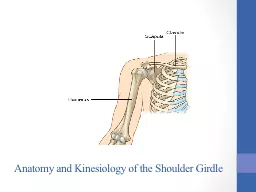
Three Bones Humerous clavicle and scapula Three Joints Glenohumeral acromioclavicular and sternoclavicular Glenohumeral Joint Acromioclavicular S ternoclavicular Rotator Cuff ID: 933659
Download Presentation The PPT/PDF document "Shoulders Shoulder anatomy" is the property of its rightful owner. Permission is granted to download and print the materials on this web site for personal, non-commercial use only, and to display it on your personal computer provided you do not modify the materials and that you retain all copyright notices contained in the materials. By downloading content from our website, you accept the terms of this agreement.


Slide1
Shoulders
Slide2Shoulder anatomy
Three Bones:
Humerous
, clavicle, and scapula
Three Joints:
Glenohumeral
,
acromioclavicular
, and
sternoclavicular
Slide3Glenohumeral Joint
Slide4Acromioclavicular
Slide5S
ternoclavicular
Slide6Rotator Cuff
What is the rotator
cuff injury?
glenohumeral
compression, rotation and dynamic stability
The Rotator cuff is made up of: supraspinatus,
infraspinatus
,
teres
minor, and
subscapularis
(SITS)
Shoulder pain is the worst pain
What type of joint is the shoulder (
glenohumeral
joint)?
Ball and socket
Slide7Function of SITS
Supraspinatous
Abduct the shoulder
Stabilize the head of
humerus
in the
glenoid
Infraspinatous
Lateral rotate
Adduct the shoulder
Stabilize the head of
humerus
in the
glenoid
Teres
Minor
Lateral rotate
Stabilize the head of
humerus
in the
glenoid
Subscapularis
Medially rotate
Stabilize the head of
humerus
in the
glenoid
.
Slide8Rotator Cuff tear/tendonitis
Degenerative process
More prevalent with advancing age
Not all RC tears require
surgury
Complete tear/partial tear usually occur with increasing age of populations
Muscle imbalance and capsular tightness impact the rotator cuff pathology and outcomes.
Slide9Treatment?
Posture reeducation
ROM
PROM,AAROM, AROM
Strengthening
Education proper positioning (Support elbow while driving).
PAMs.
Manual therapy
Slide10RC no tear/small tear
PROM and AAROM are initiated
May present with limited shoulder flexion and internal rotation.
Phase 1: forward flexion and ER supine (minimizes excessive tension)
Phase 2: extension, internal rotation and cross-body stretches.
Slide11Treatment Ideas?
Slide12Shoulder Impingements
Excessive and repetitive contact of the greater tuberosity of the humeral head with the
posterosuperior
aspect of the
glenoid
when it is repetitive abduction and external rotation.
Subscapularis
: Between the coracoid process and lesser tuberosity
Also identify as in the impingement category.
Slide13Slide14Importance of the scapula
Main stabilizers
Levator
scapula, rhomboid,
serratus
anterior, and trapezius
Improve scapular stabilization= better posture=more functional during ADL/IADL tasks.
Scapular plays major role in shoulder function.
Slide15Scapulohumeral
rhythem
First 30 degrees of shoulder abduction, the scapular remains stationary
For every two degrees of
glenohumeral
movement, for every 1 degree of
scapulothoracic
movement
Slide16Question
What nerve is involved with scapular winging:
A
.)
Thoracodorsal
nerve
B.) Axillary
C.)Long thoracic nerve
Slide17Question
What are the movements of the scapula:
Elevation/depression, protraction/retraction, IR/ER, anterior/posterior tilt.
Slide18Question
What two joint are under the most stress if a person has a hunch-over posture
SC and AC
Slide19Question
What does a
kyphotic
posture look like?
Slide20Slide21Question
What provocative test is this?
Slide22Question
What pathology does the speed’s test test for?
Long head of the biceps and superior
glenoid
labrum
Slide23Frozen Shoulder
Other name for FS
-Adhesive Capsulitis
Freezing Phase
-Pain starts with sleep and ADL tasks
-Client tends to limit movement due to an increase in pain
Frozen Phase
-This may last up to a year and compensate for GH by substituting ST motion
Thawing Phase
-Can last up to 26 months. Recent study shows 90% of patients have return full motion when compared to their contralateral side.
Slide24Thoracic Outlet Syndrome (TOS)
Compression can happen at:
Scalene triangle,
costoclavicular
space, and
pec
minor
Vascular damage is uncommon (3% to 5%)
Majority of TOS is brachial plexus related.
Slide25Question
An OTR receives an order to work on a
nonresistive
exercise program with a patient who had a shoulder fracture 2 weeks ago.
Initial
OT treatment should include:
A. pendulum
exercises
B. active
ROM
C. weight
bearing exercises
D. isotonic
strengthening
Slide26Question
An OTR is treating a patient who has a C6 complete spinal cord injury. The patient demonstrates Fair plus (3+/5) strength in scapular depression and Fair (3/5) should flexion and abduction bilaterally. The patient’s goal is to be able to sit at the edge of the bed independently. The
best
compensatory strategy for this patient to use would be full wrist extension along with shoulder:
A.)depression
, protraction, and external rotation
B.)elevation
, protraction, and external rotation
C.)elevation
, retraction, and internal rotation
D.)depression
, retraction, and internal rotation
Slide27Question
During an upper extremity assessment, a patient demonstrates 45 degrees of active shoulder flexion while in a seated position. The OTR is able to passively move the limb through the full ROM. To accurately grade the strength of this muscle group, the OTR should
NEXT
:
A.)apply
resistance in midrange with the shoulder in the frontal plane
B.)apply
resistance in midrange to the opposing muscle groups
C.)determine
the end-feel of the
glenohumeral
joint
D.)determine
active motion in a gravity-eliminated position
Slide28Question
6. A patient who had a left CVA a month ago reports constant pain in the right arm. The OTR notes that the patient’s right hand is hypersensitive and the skin is mottled. This condition is indicative of:
A.)a
hand contracture
B.)complex
regional pain syndrome
C.)a
brachial plexus injury
D.)thalamic
pain syndrome
Slide29Question
A patient who has had a CVA has mild motor and sensory loss in the upper extremity. The patient tells the OTR that the lotion being provided for sensory input is causing a skin rash. The OTRs
best
response would be to:
A.)have
the patient rinse with water after using lotion
B.)refer
the patient to an allergist
C.)use
alternating heat and cold prior to applying lotion
D.)rub
the arm with objects of varied textures instead of lotion
Slide30Question
Email:
tutorcory.passtheot@gmail.com