
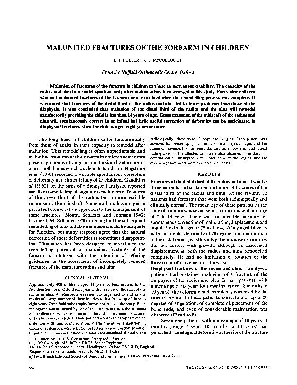
IN ADULTS 1 COLLES FRACTURE 2 SMITHS FRACTURE 3 DISTAL FOREARM FRACTURES IN CHILDREN 4 FRACTURED RADIAL STYLOID Chauffeurs fracture 5 FRACTURESUBLUXATION BARTONS FRACTURE ID: 930504
Download Presentation The PPT/PDF document "FRACTURES OF THE DISTAL RADIUS" is the property of its rightful owner. Permission is granted to download and print the materials on this web site for personal, non-commercial use only, and to display it on your personal computer provided you do not modify the materials and that you retain all copyright notices contained in the materials. By downloading content from our website, you accept the terms of this agreement.


Slide1
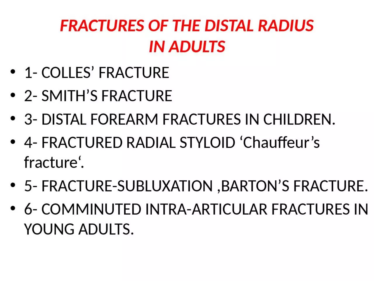
FRACTURES OF THE DISTAL RADIUSIN ADULTS
1- COLLES’ FRACTURE
2- SMITH’S FRACTURE
3- DISTAL FOREARM FRACTURES IN CHILDREN.
4- FRACTURED RADIAL STYLOID ‘Chauffeur’s fracture‘.
5- FRACTURE-SUBLUXATION ,BARTON’S FRACTURE.
6- COMMINUTED INTRA-ARTICULAR FRACTURES IN YOUNG ADULTS.
Slide2COLLES’ FRACTUREA transverse fracture of the radius just above the
wrist, with
dorsal displacement of the distal fragment
.
It
is the
most common of all fractures in
older people
,
the high
incidence being related to the onset
of
postmenopausal osteoporos
is
. Thus the patient is
usually an
older woman who gives a history of falling on
her outstretched
hand.
Slide3Clinical featuresWe can recognize this fracture (as Colles did
long before
radiography was invented) by the ‘
dinner-fork’ deformity
, with prominence on the back of the
wrist and
a depression
infront
.
In
patients with
less deformity
there may only be
local
tenderness,swelling
and
pain
on
wrist movements.
Slide4Slide5X-RAY
There is a transverse fracture of the radius at
the
corticocancellous
junction, and often the
ulnar
styloid
process
is broken off. The radial fragment is impacted into radial and backward tilt. Sometimes there is an intra-articular fracture; sometimes it is severely comminuted.
Slide6TREATMENT
1-If the fracture is
undisplaced
:
dorsal splint is applied for a day or two
untilthe
swelling has resolved, then the cast is
completed.An
x-ray is taken
at 10–14 day
s to ensure that the fracturehas not displaced; the cast can be removed
after 4 weeks to allow mobilization.2-
Displaced fractures
must be reduced under
anaesthesia
(
haematoma
block, Bier’s block or
axillary
block
). The
hand is grasped and traction is applied in
the length
of the bone (sometimes with extension of
the wrist
to
disimpact
the fragments); the distal
fragment is
then pushed into place by pressing on the
dorsum while
manipulating the wrist into flexion,
ulnar
deviation and
pronation
. The position is then checked
by x-ray
. If it is satisfactory, a dorsal plaster slab is
applied, extending
from just below the elbow to
the metacarpal
necks and two-thirds of the way round
the
circumference of the wrist. It is held in position by
a crepe
bandage.
Extreme positions of flexion and
ulnar
deviation
must be avoided; 20 degrees in each
direction
is adequate. The
arm is kept elevated for the next day or
two; shoulder
and finger exercises are started as soon
as possible
. If the fingers become swollen, cyanosed
or painful
, there should be no hesitation in splitting
the bandage. At
7–10 days fresh x-rays are taken;
re-displacement is
not uncommon and should be
treated.
Slide7The fracture unites in about 6 weeks and, even in the absence of radiological proof of union, the slab may safely be discarded and exercises
begun.
Slide8Closed reduction of Colle’s fracture
Slide9Colle’s
fracture cast
Slide103-IMPACTED OR COMMINUTED COLLES’ FRACTURES
With
substantial impaction or
comminution
in
osteoporotic bon
e
, manipulation and plaster
immobilization alone
may be insufficient. The fracture can
sometimes be reduced and held with percutaneous
wires, but if impaction is severe even this may not be enough to maintain length; in that case, an
external
fixator
is
used to
neutralize the compressive force of the 25
tendons crossing
the wrist, and bone graft or bone substitute
is placed
into the gap. The
fixator
is attached to the
distal radius
and the second metacarpal shaft.
Slide11IMPACTED OR COMMINUTED COLLES’ FRACTURES
Slide12Complications
EARLY
1-Circulatory problems
The circulation in the fingers must
be checked; the bandage holding the slab ay need to be split or loosened.
2-Nerve injury
Direct injury is rare, but compression of
the median nerve in the carpal tunnel is fairly common.
3-Reflex sympathetic
dystrophy This condition is probably
quite common, but fortunately it seldom progresses tothe full-blown picture of
Sudeck’s atrophy(swelling and tenderness and osteoporosis).4-TFCC injury TFCC injury is more common than is generally appreciated, As the distal radius displaces dorsally, the TFCC is damaged; the
ulnar
styloid
fracture
Slide13LATE1-Malunion
is common,
eitherbecause
reduction was not complete or because displacement within the plaster was overlooked.
2-Delayed union and non-union
of the radius
is rare.
3-Stiffness Stiffness of the shoulder, elbow and fingers
from neglect is a common complication. Stiffness of the wrist may follow prolonged
splintage.4-
Tendon rupture Rupture of extensor pollicis
longus
occasionally occurs a few weeks after fracture.
Slide14SMITH’S FRACTURESmith described a similar fracture about 20 years later. However, in this injury the distal fragment is displaced
anteriorly
(which is why it is sometimes called a ‘reversed
Colles
’),It is caused by a fall on the back of the hand.
Slide15Clinical featuresThe patient presents with a wrist injury, but there is no dinner-fork deformity. Instead, there is a ‘garden spade’ deformity.
Slide16X-rayThere is a fracture through the distal radial metaphysis
; a lateral view shows that the distal
fragment is displaced and tilted
anteriorly
– the
opposite of a
Colles
’ fracture.
Slide17TreatmentThe fracture is reduced by traction, supination and extension of the wrist, and the forearm is immobilized in a cast for 6 weeks. X-rays should be taken at 7–10 days to ensure the fracture has not slipped. Unstable fractures should be fixed with
percutaneous
wires or a
plate.
Slide18DISTAL FOREARM FRACTURES INCHILDREN
The distal radius and ulna are among the commonest sites of childhood fractures. The break may occur through the distal radial
physis
or in the
metaphysis
of one or both bones.
Metaphyseal
fractures are often incomplete or greenstick
Mechanism of injury
The usual injury is a fall on the outstretched hand with the wrist in extension; the distal fragment is forced
posteriorly (this is often called
a ‘juvenile Colles’ fracture’).
Slide19Clinical features
There is usually a history of a fall, though this may be passed off as one of many childhood spills. The wrist is painful, and often quite swollen; sometimes there is an obvious
‘dinner-fork
’ deformity.
X-ray
The precise diagnosis is made on the x-ray
appearances.
Physeal
fractures are almost
invariably Salter–Harris
type I or II, with the epiphysis shifted and tilted backwards and
radially. Type V injuries are unusual;sometimes they are diagnosed in retrospect when
premature
epiphyseal
fusion occurs.
Metaphyseal
injuries may appear as mere buckling of
the cortex.
Slide20TreatmentPhyseal fractures are reduced, under
anaesthesia
, by
pressure on the distal fragment. The arm is immobilized in a full-length cast with the wrist slightly flexed and
ulnar
deviated, and the elbow at 90 degrees. The cast is retained for 4-6 weeks.
If the fracture slips, especially if the ulna is intact, it should be stabilized with a
percutaneous
K-wire.
Slide21FRACTURED RADIAL STYLOIDThis injury is caused by forced radial deviation of the wrist and may occur after a fall, or when a starting handle ‘kicks back’ – the so-called ‘
chauffeur’s
fracture‘
The
fracture line is transverse, extending laterally from the
articular
surface of the radius; the fragment, much more than the radial
styloid
, is often
undisplaced
.
Slide22Treatment
If there is displacement it is reduced, and the wrist is held in
ulnar
deviation by a plaster slab round the outer forearm extending from below the elbow to the metacarpal necks. Imperfect reduction may lead to osteoarthritis; therefore if closed reduction is imperfect the fragment should be screwed back, or held with K-wires.
Slide23Slide24FRACTURE-SUBLUXATION (BARTON’SFRACTURE)1-volar barton
2-dorsal
barton
Slide25COMMINUTED INTRA-ARTICULARFRACTURES IN YOUNG ADULTS
In the young adult, a comminuted intra-
articular
fracture
is a high energy injury. A poor outcome
will result
unless intra-
articular
congruity, fracture
alignment and
length are restored and movements started as soon as possible. For these patients a much higher standard must be set than would be accepted for the typical
osteoporotic fracture. In addition to the usual posteroanterior and lateral x-rays, oblique views and often
CT scans are useful to show the fragment
alignment. The
simplest option is a manipulation and cast.
If the
anatomy is not restored, then an open
reduction
may be necessary. The medial complex must
be anatomically
reduced, which may require open
reduction through
dorsal and
palmar
approaches and
a combination
of wires, plates, screws and bone
grafts.
Slide26Slide27CARPAL BONES
Slide28FRACTURED SCAPHOID
Scaphoid
fractures account for almost 75 per cent of all carpal fractures although they are rare in the elderly and in children.
Slide29Mechanism of injury and pathological anatomy
The
scaphoid
lies obliquely across the two rows of carpal bones, and is also in the line of loading between
the thumb and forearm. The combination of forced
carpal movement and compression, as in a fall on the
dorsiflexed
hand, exerts severe stress on the bone and
it is liable to fracture. Most
scaphoid
fractures are stable;with unstable fractures the fragments may become displaced.
Slide30Clinical features
The appearance may be normal, but there is can usually
detect fullness in the anatomical snuffbox
; precisely localized
tenderness
in the same place is an important diagnostic sign; the
scaphoid
can of course also be palpated from the front
and back of the wrist and it may be tender there as well. Proximal pressure along the axis of the thumb may be
painful
.
Slide31X-ray
Anteroposterior
, lateral and oblique views are all essential; often a recent fracture shows only in the oblique view. Usually the fracture line is
transverse,and
through the narrowest part of the bone (waist), but it may be more proximally situated (proximal pole fracture). Sometimes only the tubercle of the
scaphoid
is fracture
d
.
Slide32Treatment
1-Undisplaced fractures
need no reduction and are
treated in plaster. The cast is applied from the upper forearm to just short of the
metacarpo-phalangeal
joints of the fingers, but incorporating the proximal phalanx of the thumb. The wrist is held
dorsiflexed
and the thumb forwards in the
‘glass-holding’ position
. It is retained for
8 weeks
. After 8 weeks the plaster is removed and the wrist examined clinically and radiologically. If there is no tenderness and the x-ray shows signs of healing, the wrist is left free; a CT scan is the most reliable means of confirming union if in doubt.
If the
scaphoid
is tender, or the fracture still visible on x-ray, the cast is reapplied for a further 4 weeks.
After that either the fracture united with pain free or end with non union and in this case need bone graft and internal fixation by
herbert
screw.
Slide33Scaphoid frcature
Slide342-Displaced fractures can also be treated in plaster, but the outcome is less predictable. It is better to reduce the fracture openly and to fix it with a compression screw.
Slide35Complications
1-Avascular necrosis
The proximal fragment may
die,
especially
with proximal pole fractures, and then at 2–3 months it appears dense on x-ray.
Slide362-Non-union
By 3 months it may be obvious that the
fracture will not unite with sclerosis and
cavitation
. Bone grafting with compression screw should be attempted, especially in the younger, more vigorous type of patient, because this probably reduces the chance of later, symptomatic osteoarthritis.
Slide373-Osteoarthritis:
Non-union or
avascular
necrosis may
lead to secondary osteoarthritis of the wrist.
The treatment either by wrist
arthrodesis
or proximal raw
carpectomy
.
Slide38