

Most of these lesions are benign Breast cancer is 2 nd most common cause of cancer deaths in women following carcinoma of the lung The clinical significance of the benign conditions 1 possible clinical confusion with malignancy ID: 934603
Download Presentation The PPT/PDF document "Pathology of The Breast Lesions of femal..." is the property of its rightful owner. Permission is granted to download and print the materials on this web site for personal, non-commercial use only, and to display it on your personal computer provided you do not modify the materials and that you retain all copyright notices contained in the materials. By downloading content from our website, you accept the terms of this agreement.


Slide1
Pathology of The Breast
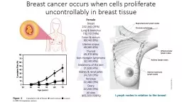
Slide2Lesions of female breast are much more common than lesions of male breastMost of these lesions are benign
Breast cancer is 2
nd
most common cause of cancer deaths in women, following
carcinoma of the lung.
The clinical significance of the
benign
conditions:
1- possible clinical confusion with malignancy
2- association of certain variants with breast carcinoma.
Slide3Breast diseases
Slide4Fibrocystic changes
are the most common cause of breast "lumps
exaggeration and distortion of the cyclic breast changes that occur normally in the menstrual cycle
.
HRT and OCPs do
not
increase the incidence of these alterations; (OCPs may
decrease
the risk).
arise during reproductive period of life
Very common (at autopsy in 60% to 80% of women)
Slide5TUMORS OF THE BREAST
Slide61-
Fibroadenoma
The most common benign neoplasm of the female breast.
increase in estrogen activity
Most in third decade of life.
a discrete, solitary, freely movable nodule, (1 to 10 cm).
usually easily "shelled out“ surgically.
may enlarge late in the menstrual cycle and during pregnancy.
After menopause they usually regress and calcify.
Slide7Cytogenetic studies stromal
cells are monoclonal and so represent the
neoplastic
element of these tumors (
the
neoplastic
stromal
cells secrete growth factors that induce proliferation of epithelial cells
).
Fibroadenomas almost never become malignant.
Fibroadenoma
Slide8Fibroadenoma
Slide9Phyllodes
Tumor
much less common than
fibroadenomas
arise from the
periductal
stroma
and
not
from preexisting
fibroadenomas
.
leaflike
clefts and slits
they have been designated
phyllodes
(Greek for "
leaflike
")
Most (70% )are benign and tend to remain localized and cured by excision.
The most worrying change
the appearance of increased
stromal
cellularity
with
anaplasia
and high
mitotic activity
, accompanied by rapid increase in size, usually with
invasion
of adjacent breast tissue =
malignant
phyllodes
.
Malignant lesions may recur
15% of cases
metastasize to distant sites.
Slide10Carcinoma of the Breast
the most common cancer in females
ranking second
only to lung cancer as a cause of
cancer
death
in women.
75% of women with breast cancer are
older than age 50
.
Only 5% are younger than the age of 40.
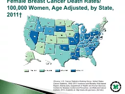
Slide11CA: A Cancer Journal for Clinicians
Volume 62, Issue 1,
pages 10-29, 4 JAN 2012 DOI: 10.3322/caac.20138
http://onlinelibrary.wiley.com/doi/10.3322/caac.20138/full#fig1
Slide12Pathogenesis
(1)Genetic Changes
well-established familial syndromes
-sporadic breast cancer:
e.g.
overexpression
of the
HER2/NEU
proto-oncogene
(30% of cases)
(2)Hormonal Influences
increased exposure to
estrogen
(3)Environmental Variables
Slide13Factor
Relative Risk
Well-Established Influences
Geographic factors
Varies in different areas
Age
Increases after age 30yr
Family history
First-degree relative with breast cancer
1.2-3.0
Premenopausal
3.1
Premenopausal and bilateral
8.5-9.0
Postmenopausal
1.5
Postmenopausal and bilateral
4.0-5.4
Menstrual history
Age at menarche <12yr
1.3
Age at menopause >55yr
1.5-2.0
Pregnancy
First live birth from ages 25 to 29yr
1.5 First live birth after age 30yr1.9 First live birth after age 35yr2.0-3.0 Nulliparous3.0Benign breast disease Proliferative disease without atypia1.6 Proliferative disease with atypical hyperplasia>2.0 Lobular carcinoma in situ6.9-12.0Less Well-Established Influences Exogenous estrogens Oral contraceptives Obesity High-fat diet Alcohol consumption Cigarette smoking
Slide14Major Risk Factors
Age.
Genetics and Family History:
50% of women with
hereditary
breast cancer have mutations in gene
BRCA1
; 30%
have mutations in
BRCA2
.
other genetic diseases may be associated with breast cancer
Prolonged exposure to exogenous estrogens
postmenopausally
(HRT)
Ionizing radiation, in early life years
Slide15Morphology of breast cancer
About 4% of cases
bilateral primary tumors or sequential lesions in the same breast.
- The locations of the tumors within the breast are:
Upper outer quadrant 50% (most common)
Central portion 20%
Lower outer quadrant 10%
Upper inner quadrant 10%
Lower inner quadrant 10%
Slide16Breast cancers are classified into:Noninvasive
(
confined by a basement membrane and do not invade
into
stroma
or
lymphovascular
channels), include:
Ductal carcinoma in situ (DCIS)
Lobular carcinoma in situ (LCIS)
Invasive (infiltrating)Invasive ductal carcinoma – NOS (most common type)Invasive lobular carcinoma
Medullary carcinoma
Colloid (mucinous) carcinoma
Tubular carcinoma
Other types
Slide17Slide18Ductal carcinoma in-situ DCIS
ranges from low nuclear grade to pleomorphic (high nuclear grade).
comedo
subtype: high-grade nuclei with
extensive central necrosis
. (The name derives from the toothpaste-like necrotic tissue).
Calcifications
are frequently
associated
screening
by mammography
The prognosis : excellent (97% long-term survival
afte
r simple mastectomy)
Current treatment strategies: surgery and radiation,
tamoxifen
Significance: adjacent invasive CA; become invasive if untreated
Slide19Comedo DCIS
Slide20Invasive ductal
carcinoma
Also called
Carcinomas "not otherwise specified"
70% to 80% of all
Precancerous lesion
: usually DCIS
Clinical presentation:
a mammographic density; a hard, palpable mass. Advanced cancers may cause retraction of the nipple, or fixation to the chest wall.
Receptor profile
: 2/3 express ER or PR; 1/3
overexpresses
HER2/NEU.
Slide21Invasive
ductal
carcinoma
Slide22Invasive lobular carcinoma
These tumors comprise fewer than
20%
of all breast carcinomas.
Precancerous
lesion
. 2/3 adjacent LCIS.
multicentric
and bilateral (10% to 20%).
Clinical presentation
. Most present as palpable masses or mammographic densities
Almost all of these carcinomas express hormone receptors, but HER2/NEU
overexpression
is very rare or absent.
Slide23Medullary carcinoma
is a rare subtype
1%
of cases.
Microscopically:
large
anaplastic
cells with pushing, well-circumscribed borders. With a pronounced
lymphoplasmacytic
infiltrate.
Precancerous
lesions
. usually absent
increased frequency in women with
BRCA1
mutations
,.
Receptor
profile
. lack hormone receptors and do not overexpress HER2/NEU.
Slide24Slide25Colloid (mucinous) carcinoma
a rare subtype.
Microscopic
picture
. The tumor cells produce abundant quantities of extracellular
mucin
that dissects into the surrounding
stroma
. Grossly the tumors are usually soft and gelatinous.
Most express hormone receptors (ER,PR), and rare examples may
overexpress
HER2/NEU.
Slide26Slide27Tubular carcinomas
10%
of invasive carcinomas smaller than 1 cm found with mammographic screening.
Clinical
presentation
. irregular mammographic densities.
Microscopically
, well-formed tubules with low-grade nuclei.
Lymph node metastases are rare, and prognosis is excellent.
Virtually all tubular carcinomas express hormone receptors, but
overexpression
of HER2/NEU is uncommon.
Slide28Slide29Features Common to All Invasive Cancers
Fixation:
adherent to the pectoral muscles or deep fascia of the chest wall
retraction
or
dimpling
of the skin or nipple: adherence to the overlying skin
peau
d'orange (orange peel): Involvement of the lymphatic pathways cause localized
lymphedema
, the skin becomes thickened around exaggerated hair follicles
Slide30Spread of Breast Cancer
through
lymphatic
and
hematogenous
channels.
Favored
mets
are the
lungs
, skeleton,
liver
, and
adrenals
and (less commonly) the brain, spleen, and pituitary.
Metastases may appear many years after apparent therapeutic control of the primary lesion
SCREENING
:
mammographic screening
Magnetic resonance imaging MRI
Slide31Prognosis
1-
The size
.
2-
Lymph node involvement and the number of lymph nodes involved by metastases
.
3-
Distant
metastases.4- grade5- The
histologic
type of carcinoma
6- The presence or absence of estrogen or progesterone receptors
.
7-
The proliferative rate of the cancer
.
8-
Aneuploidy
.
worse prognosis.
9- Overexpression of HER2/NEUthe importance of evaluating HER2/NEU is to predict response to a monoclonal antibody ("Herceptin
") against the gene product.
Slide32Male breast pathology
Gynecomastia
Enlargement of the male breast
due to absolute or relative estrogen excesses.
According to cause, divided into:
1-
pathologic
gynecomastia
: cirrhosis of the liver;
Klinefelter
syndrome; estrogen-secreting tumors; estrogen therapy; digitalis therapy.
2- Physiologic
gynecomastia
:
puberty and extreme old age.
Slide33Carcinoma of the male breast
male: female breast cancer
1: 125.
advanced age.
Because of the scant amount of breast substance in the male, the tumor rapidly infiltrates the overlying skin and underlying thoracic wall.
Unfortunately, almost
1/2
have spread to regional lymph nodes and more distant sites by the time they are discovered.