
Content Ryan Martin MD Sarah Peacock DNP APRN ACNPBC Megan Corry EdD EMTP Kerri L LaRovere MD Safdar A Ansari MD Slides Ryan Martin MD Presenter Your name ID: 929979
Download Presentation The PPT/PDF document "ENLS Version 4.0 Spinal Cord Compression" is the property of its rightful owner. Permission is granted to download and print the materials on this web site for personal, non-commercial use only, and to display it on your personal computer provided you do not modify the materials and that you retain all copyright notices contained in the materials. By downloading content from our website, you accept the terms of this agreement.


Slide1
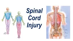
Slide2ENLS Version 4.0Spinal Cord Compression
Content: Ryan Martin, MD; Sarah Peacock, DNP, APRN. ACNP-BC; Megan Corry, EdD, EMTP; Kerri L. LaRovere, MD; Safdar A. Ansari, MD Slides: Ryan Martin, MD
Slide3Presenter:Your name
Conflicts:
No conflicts
Slide4Editors’ Note: Global Considerations
The intent of the editors, authors, and reviewers of this ENLS topic was not to address all the variations in international practice for the different diseases. We have discussed major practice variances (e.g., the availability of diagnostic testing, or the type of medications used) and encourage learners to use the ENLS algorithms as a framework on which any relevant local practice guidelines can be incorporated.
Slide5Slide6ENLS: Spinal Cord Compression
Learning Objectives:Recognize the signs and symptoms of acute myelopathyDescribe the role of imaging in the management of spinal cord compressionDescribe empirical treatments for neoplastic-associated myelopathy and infection-related myelopathy
Slide7Checklist
☐ Brief history of the patient
☐
Spinal motion restriction (motion restriction)
☐ Ensure proper ventilation, especially in the presence of quadriplegia
☐ Laboratories: CBC, chemistries, INR/PT, PTT, platelet function assay (e.g.,
VerifyNow
platelet reactivity profile)
☐ Obtain emergent spine imaging (MRI unless contraindicated)
☐ Alert spine surgeon
☐ Suspected neoplastic disease: administer corticosteroids, contact radiation oncology
☐ Suspect epidural infection: check ESR and start antibiotics
☐ Initiate interfacility transfer if anything cannot be performed at your facility
Slide8Myelopathy Presentation
Spinal Cord Compression
Neurological dysfunction at or below the level of compression
Most common causes:
Trauma
Malignancy
Degenerative Spine Disease
Infection
Hematoma
Neurological dysfunction
Weakness
Paraplegia or quadriplegia
Acute or progressive
Sensory changes
Determine the level
Sphincter dysfunction
Consider cervical collar
Slide9Airway and HemodynamicsMust evaluate, especially if the patient is quadriplegic
Assessment of respiratory functionMIF/NIF, VC Acute loss of sympathetic toneNeurogenic shock; treat with fluids, pressors
Slide10ImagingMRI
Modality of choiceSoft tissues and discrete regionsExtradural, intradural, intramedullaryLong segments imaged
CT or CT Myelography
CT is standard scan if trauma is suspected
May be useful in patients with MRI contraindications
Slide11Emergent TransferTransfer if your facility is unable to provide definitive imaging or care
Transfer agreementsminimize prolonged transfer timesPhysician-to-physician consultation and nurse-to-nurse report is essentialEmpirical therapy should be considered if there is a delay in transfer or lengthy transport is anticipated, even if the diagnosis is not confirmed
Slide12Empirical Therapy When Imaging is
NOT available
Slide13MRI Spine Available
Slide14Case #1
75-year-old male history of prostate cancer presents with two days of lower extremity weakness, and decreased grip strengthHe has had neck pain for three weeksAn MRI of his cervical spine is obtained
Slide15Case #1
MRI confirms metastatic disease to the C7 vertebral body with spread into the spinal canal.
What is the next best step in management of this patient?
Radiation TherapyChemotherapy
CorticosteroidsConsult Spine SurgeonC and D
Slide16Case #1
MRI confirms metastatic disease to the C7 vertebral body with spread into the spinal canal.
What is the next best step in management of this patient?
Radiation TherapyChemotherapy
CorticosteroidsConsult Spine Surgeon
C and D
Slide17Metastatic Disease of Spine
Motor function predictive of outcomeSCC presenting symptom of CA 20%Extradural tumor masses present in:Thoracic spine (60%)Lumbosacral spine (30%)Cervical spine (10%)
Slide18Metastatic Disease of SpineMost common neoplasmsLung
BreastProstate Renal cell carcinomaLymphomaContiguous spread from paraspinal tumors occurs less often (15-20% of metastatic lesions)Metastases can occur to the intramedullary space
Slide19Cranial and Spinal Nerve InvolvementEarly cranial or spinal nerve dysfunction is suggestive of leptomeningeal metastases
Symptoms may include motor or sensory deficits in several non-contiguous sites Portends a poor prognosis
Slide20PlanSurgical Evaluation
CorticosteroidsEvaluation for radiotherapy / chemotherapyPain control
Slide21Case #2
33-year-old female with a history of intravenous drug abuse presents after a ground level fall. She is weak in all four extremities. She is intubated given respiratory distress.A CT of her cervical spine is performed, followed by an MRI of her cervical spine.
Slide22Case #2
CT shows severe bony erosion at C3, and her MRI shows multilevel spinal canal degenerative changes and an epidural abscess. What is the next best step in management of this patient?Consult spine surgeonIntravenous antibioticsCorticosteroidsESR
A, B, and D
Slide23Case #2
CT shows severe bony erosion at C3, and her MRI shows multilevel spinal canal degenerative changes and an epidural abscess. What is the next best step in management of this patient?Consult spine surgeonIntravenous antibioticsCorticosteroidsESR
A, B, and D
Slide24Infection
Triad: pain, fever, neurologic deficit
Rare
Hematogenous spread
Risk factors
Staphylococcus aureus
Most common
Slide25Infection Injury to the spinal cord through direct compression and by vascular compromise
Diagnosis is often delayed due to a lack of symptoms other than painDifficult to distinguish from leptomeningeal metastasesImaging with abnormalities involving two or more vertebral bodies across a disk space suggests an infection
Slide26Plan
Multi-microbial antibiotic therapyVancomycinThird or fourth generation cephalosporinLaboratory studiesBlood cultures, ESRSurgical Evaluation
Lateral c-spine x-ray
Slide27Case #3
75-year-old male with a history of diabetes and atrial fibrillation on apixaban presents with acute onset numbness and weakness in his legs, as well as mild numbness in his arms.An MRI of his cervical spine is performed.
Slide28Case #3
An epidural hematoma is noted on MRI. What is the next best step in management of this patient?Physical therapyCorticosteroidsNeurology ConsultationLumbar PunctureReverse coagulopathy
Slide29Case #3
An epidural hematoma is noted on MRI. What is the next best step in management of this patient?Physical therapyCorticosteroidsNeurology ConsultationLumbar Puncture
Reverse coagulopathy
Slide30Epidural HematomaVascular malformation
CoagulopathyMyelitisSpinal tumorsSyringomyelia
Slide31PlanSurgical Evaluation
Treat underlying causeTiming variable for surgery
Correct Coagulopathy
See ENLS ICH protocol
Slide32Case #4
A 45-year-old male presents after developing sudden onset pain radiating from his neck down his right arm. Some tingling noted in his legs as well. This occurred while lifting heavy weights at the gym. An MRI of his cervical spine is performed
Slide33Case #4
Acute Disc HerniationDisc disease usually causes radiculopathyAcute myelopathy rareCord compressionImpaired blood supplyPain
Slide34Plan Surgical Evaluation, Decompression
No consensusConsider SteroidsAnalgesia
Slide35Intrinsic lesions of the spinal cord
Slide36Intrinsic lesions of the spinal cord
Spinal Cord InfarctionMyelitisInfectiousInflammatory
Slide37Pediatric ConsiderationsGenetic conditionsMultidisciplinary approach to the patient
Cervical spine considerationsCardiovascular considerationsBradycardia MAP goal > 50th percentile for age
Slide38Communication
☐ Age, gender, pre-morbid conditions
☐ Onset and duration of symptoms
☐ Clinical spinal level of pathology
☐ Airway status and vitals
☐ Bowel or bladder involvement
☐ Results lab tests and spinal imaging
☐ What therapy has been
started
☐ Inquire
what further therapy to start now
Slide39Clinical PearlsSCC is acute neurological dysfunction below the level of compression.
Common etiologies are trauma, malignancy, degenerative spinal disease, epidural abscess, and hematomas.Quadriplegia should prompt assessment of respiratory function to detect impending respiratory failure.Empirical treatment for infectious or malignant causes of SCC is recommended if delay in MRI imaging is unavoidable.Early decompressive surgery is recommended and correlates with improved outcomes.
Slide40Questions?