
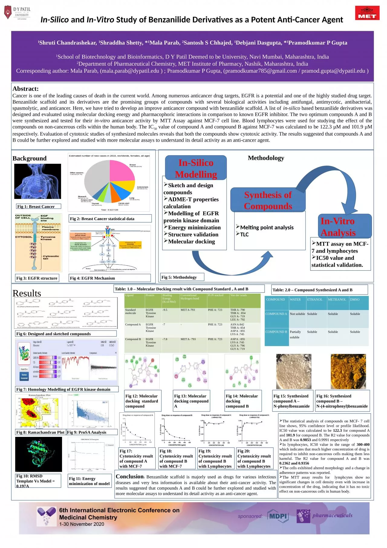
Study of Benzanilide Derivatives as a Potent AntiCancer Agent 1 Shruti Chandrashekar 1 Shraddha Shetty 1 Mala Parab 2 Santosh S Chhajed 1 Debjani Dasgupta 1 ID: 929887
Download Presentation The PPT/PDF document "In-Silico and In-Vitro" is the property of its rightful owner. Permission is granted to download and print the materials on this web site for personal, non-commercial use only, and to display it on your personal computer provided you do not modify the materials and that you retain all copyright notices contained in the materials. By downloading content from our website, you accept the terms of this agreement.


Slide1
In-Silico
and In-Vitro Study of Benzanilide Derivatives as a Potent Anti-Cancer Agent
1Shruti Chandrashekar, 1Shraddha Shetty, *1Mala Parab, 2Santosh S Chhajed, 1Debjani Dasgupta, *1Pramodkumar P Gupta1School of Biotechnology and Bioinformatics, D Y Patil Deemed to be University, Navi Mumbai, Maharashtra, India2Department of Pharmaceutical Chemistry, MET Institute of Pharmacy, Nashik, Maharashtra, IndiaCorresponding author: Mala Parab, (mala.parab@dypatil.edu ) ; Pramodkumar P Gupta, (pramodkumar785@gmail.com / pramod.gupta@dypatil.edu )
Abstract:
Cancer is one of the leading causes of death in the current world. Among numerous anticancer drug targets, EGFR is a potential and one of the highly studied drug target. Benzanilide scaffold and its derivatives are the promising groups of compounds with several biological activities including antifungal, antimycotic, antibacterial, spasmolytic, and anticancer. Here, we have tried to develop an improve anticancer compound with benzanilide scaffold. A list of in-silico based benzanilide derivatives was designed and evaluated using molecular docking energy and pharmacophoric interactions in comparison to known EGFR inhibitor. The two optimum compounds A and B were synthesized and tested for their in-vitro anticancer activity by MTT Assay against MCF-7 cell line. Blood lymphocytes were used for studying the effect of the compounds on non-cancerous cells within the human body. The IC50 value of compound A and compound B against MCF-7 was calculated to be 122.3 µM and 101.9 µM respectively. Evaluation of cytotoxic studies of synthesized molecules reveals that both the compounds show cytotoxic activity. The results suggested that compounds A and B could be further explored and studied with more molecular assays to understand its detail activity as an anti-cancer agent.
Background
Fig 6:
Designed and sketched compounds
Methodology
Sketch and design compounds
ADME-T properties calculation
Modelling of EGFR protein kinase domain
Energy minimizationStructure validationMolecular docking
In-Silico Modelling
Synthesis of Compounds
Melting point analysisTLC
In-Vitro Analysis
MTT assay on MCF-7 and lymphocytesIC50 value and statistical validation.
Results
Ligand
Protein
Binding Energy (Kcal/Mol)
Conventional Hydrogen bond
Pi
-Pi stacked
Van der waals Standard moleculeEGFR Tyrosine Kinase-9.5MET A-793PHE A: 723THR A -790THR A : 854GLY A- 719LEU A- 792Compound AEGFR Tyrosine Kinase-7-PHE A: 723ASN A:842THR A: 854ASP A : 855LYS A :745Compound BEGFR Tyrosine Kinase-7.8MET A- 793 PHE A: 723 ASP A : 855LYS A :745GLY A: 796GLY A: 719
COMPOUND
WATER
ETHANOL
METHANOL
DMSOCOMPOUND ANot solubleSolubleSolubleSolubleCOMPOUND BPartially soluble SolubleSolubleSoluble
Fig 1:
Breast Cancer
Fig 2:
Breast Cancer statistical data
Fig 3: EGFR structure
Fig 4: EGFR Mechanism
Fig 12: Molecular docking standard compound
Fig 13: Molecular docking compound A
Fig 14: Molecular docking compound B
Fig 15: Synthesized compound A – N-phenylbenzamide
Fig 16: Synthesized compound B – N-(4-nitrophenyl)benzamide
Fig 7: Homology Modelling of EGFR kinase domain
Fig 8: Ramachandran Plot
Fig 9: ProSA Analysis
Fig 10: RMSD Template Vs Model = 0.197A
Fig 11: Energy minimization of model
Table: 1.0 – Molecular Docking result with Compound Standard , A and B
Table: 2.0 – Compound Synthesized A and B
Fig 17: Cytotoxicity result of compound A with MCF-7
Fig 18: Cytotoxicity result of compound B with MCF-7
Fig 20: Cytotoxicity result of compound B with Lymphocytes
Fig 19: Cytotoxicity result of compound B with Lymphocytes
The statistical analysis of compounds on MCF- 7 cell line shows, 95% confidence level or profile likelihood. IC50 value was calculated to be
122.3
for compound A and
101.9 for compound B. The R2 value for compounds A and B was 0.9853 and 0.9991 respectivelyIn lymphocytes, IC50 value in the range of 300-400 which indicates that much higher concentration of drug is required to inhibit non-cancerous cells making them less harmful. The R2 value for compound A and B was 0.2362 and 0.9356The cells exhibited altered morphology and a change in adherence patterns was reported. The MTT assay results for lymphcytes show no significant changes in cell density even with increase in concentration of the drug, indicating that it has no toxic effect on non-cancerous cells in human body.
Conclusion: Benzanilide scaffold is majorly used as drugs for various infectious diseases and very less information is available about their anti-cancer activity. The results suggested that compounds A and B could be further explored and studied with more molecular assays to understand its detail activity as an anti-cancer agent.
Fig 5: Methodology