/
Aarti Ragoonath, Lukas Bijaminas, Aarti Ragoonath, Lukas Bijaminas,

Aarti Ragoonath, Lukas Bijaminas, - PowerPoint Presentation

RefreshingView
RefreshingView . @RefreshingView
Follow
344 views
Uploaded On 2022-08-03

Aarti Ragoonath, Lukas Bijaminas, - PPT Presentation

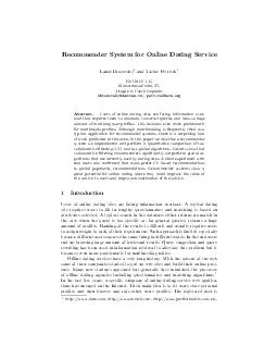
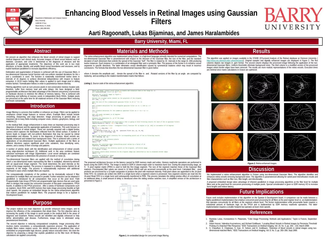
and James Haralambides Department of Mathematics and Computer Science Barry University 11300 NE 2 nd Ave Miami Shores FL 33161 Phone 305 8993035 We present an algorithm that enhances the blood vessels of retinal images to support medical diagnosis and clinical study Accurate i ID: 933348

image filter filters gaussian filter image gaussian filters pixel images dimensional retina vessels algorithm memory level multiple retinal processing

Share:

Link:

Embed:

Download Presentation from below link

Download Presentation The PPT/PDF document "Aarti Ragoonath, Lukas Bijaminas," is the property of its rightful owner. Permission is granted to download and print the materials on this web site for personal, non-commercial use only, and to display it on your personal computer provided you do not modify the materials and that you retain all copyright notices contained in the materials. By downloading content from our website, you accept the terms of this agreement.


Presentation Transcript

Slide1

Aarti Ragoonath, Lukas Bijaminas,

and James Haralambides

Department of Mathematics and Computer Science Barry University11300 NE 2nd Ave.Miami Shores, FL 33161Phone: (305) 899-3035

We present an algorithm that enhances the blood vessels of retinal images to support medical diagnosis and clinical study. Accurate imagery of blood vessel features such as diameter, curvature, and color is detrimental to the diagnosis of diseases and the application of appropriate treatments. The objectives of this work are in two main directions: a) locate, identify, and amplify blood vessel boundaries and structures, and b) exploit hardware parallelism to increase algorithmic efficiency. Delineation and augmentation of features is achieved with the use of Gaussian filters. A two-dimensional Gaussian kernel function with non-uniform standard deviations for the x and y coordinates is used. The function is rotationally transformed twelve times in intervals of 15 degrees to achieve directional independence with respect to feature extraction. A 1515 matrix holding filter values is applied to each image pixel in sliding window fashion. Normalization follows in order to parameterize feature intensity.Filtering algorithms such as ours process pixels in non-consecutive memory location and, therefore, suffer from memory read and write delays. We have designed a field-programmable architecture that takes advantage of the inherent concurrency of processes on hardware devices to minimize the effects of memory latency. This is achieved with prefetching and buffering of memory words in independent block RAMs. Multiple pixels are processed at the same time for all rotational versions of the Gaussian filters reducing overheads substantially.

Enhancing Blood Vessels in Retinal Images using Gaussian Filters

H

2O

Abstract

Introduction

References

Barry University, Miami,

FL

Rastislav Lukac, Konstantinos

N. Plataniotis, “Color Image Processing: Methods and Applications,” Taylor & Francis, September 2006.Adam Hoover, Valentina Kouznetsova, and Michael Goldbaum, “Locating Blood Vessels in Retinal Images by Piecewise Threshold Probing of a Matched Filter Response,” IEEE Transactions on Medical Imaging, Vol. 19, No. 3, pp. 203 – 210, March 2000.S. Chaudhuri, S. Chatterjee, N. Katz, M. Nelson, and M. Goldbaum, “Detection of blood vessels in retinal images using two-dimensional matched filters,” IEEE Transactions on Medical Imaging, Vol. 8, No. 3, pp. 263–269, Sept.1989.

Materials and Methods

Results

Discussion

Future Implications

Purpose

The retina enhancement algorithm

was implemented using Octave, is a high-level language primarily intended for numerical computations. The two-dimensional Gaussian filter is parameterized with respect to the direction of the Gaussian filter, the size of the filter, and the standard deviation of each dimension that controls the spread of the Gaussian “bell”. The filter is rotated by 15 intervals in the range [0, 180) producing twelve instances. Each instance is a combination of an elongated filter and a symmetric filter. The purpose of the former is to enhance vessel segments in specific directions. The latter eliminates vessel identification caused by symmetrical rotations which may result in incorrect conclusions. The formula that describes the two-dimensional Gaussian filter is given below:, where A denotes the amplitude and , denote the spread of the filter in and . Rotated versions of the filter by an angle are computed by replacing and according to the rotation transformation matrix that follows:Listing 1. Source code of the retina enhancement algorithm.


Image filtering is a process that modifies or enhances an image. Filters can be applied to emphasize certain image features or remove others. Example filters include include smoothing, sharpening, and edge detection. Image processing in general plays an important role in many fields including computer vision, robotics, geophysics, biology, and medicine [1].In the medical field, image enhancement is many times an important processing step in diagnosis of disease and the appropriate application of treatments. This work focuses on the enhancement of retinal images. These are normally acquired with a digital fundus camera which captures the illumination reflected from the retinal surface. A number of factors leads to images with non-uniform illumination that may be caused by retinal abnormalities and disease. To assist in the diagnosis of disease, blood vessels are digitally enhanced and isolated from background noise. This is achieved with a series of applications of two-dimensional Gaussian filters. Multiple applications of the filters at different directions capture significant pixel color variations, thus identifying veins, arteries, and a variety of their crossings and patterns.A number of articles deals with the identification and enhancement of retinal vessels using edge-detection techniques [2]. Additional work in the area combines feature extraction techniques with image segmentation. Local vessel attributes are combined with regional features to determines the presence of vessels or noise [3].Two-dimensional Gaussian filters are applied with the method of convolution during which a two-dimensional matrix representing the filter is multiplied, element-by-element with an equal-sized image segment. The result determines the pixel intensity at the center of the image segment. The process is repeated in a sliding window fashion for all segments of the image. The process is computationally expensive with additional overhead in cases where multiple filters are required.The computationally complexity of the problem can be dramatically reduced if filter operations for multiple filters are executed in parallel. Further reduction can be obtained if parallelism is introduced for all computations that occur at the pixel level. Field Programmable Gate Arrays (FPGAs) are programmable logic devices that are used quite often in designs that require the concurrent execution of fine-grain computations. FPGA boards, in addition to the FPGA processor, offer a variety of hardware components such as registers, block RAM, and DDR memory that make image processing feasible at high clock speeds. We propose an embedded design for the retina enhancement algorithm that realizes parallelism for multiple filters. The proposed design is for a Spartan 6 Development board.

The project realizes two main objectives: a) provide enhanced retina images, and b) improve the efficiency of the computations that obtain them. The first objective aims at increasing the quality of the image to assist people in the medical field in the areas of diagnosis and treatment. Retina vessels are identified and digitally enhanced to help define abnormalities and disease. Furthermore, image noise is eliminated using parameterized thresholds.The second objective is reducing computational complexity. Image processing often involves matrix convolution, a computationally expensive process. The application of multiple filters makes matters worse. We identify elements of parallelism that, when embedded on programmable logic devices, greatly reduce execution times. We meet the objective by proposing a design that exploits parallelism at the filter level. All filtering calculations are applied concurrently.

%Read the image, convert pixel values to double and normalize them in the range [0, 1]im = double(imread('im0001.ppm')) / 255;%Display the original image in figure 1figure(1), imshow(im)%Select the stronger green channel as the grayscale of the originalim = im(:, :, 2);%Set the size of the 2-dimensional gaussian filter to 23 (2 * N + 1)N=11;%Set standard deviations for the elongated gaussian filter as sx = 1 and sy = 4sx = 1;sy = 4;%Set standard deviation for the symmetric gaussian filter as s = sx = sy = 4s = sy;%Set the angle increment to 15 degreesang_step = 15;%Compute the dimensions [x, y] of the imageims = size(im);%Construct a 3-dimensional all-zeros matrix that is going to hold filtering results for each of the 12 gaussian filtersimg=zeros(ims(1), ims(2), 180 / ang_step);%Apply the (symmetric filter - elongated filter) function for 12 angles in [0, 165] in increments of 15 degreesk = 0;for th = 0 : ang_step : 165 k = k + 1; g = gaussian_filter (s, s, th, N) - gaussian_filter (sx, sy, th, N); img(:, :, k) = filter2(g, im);end%Select the maximum pixel intensity for corresponding pixels out of the 12 filtering resultsimm = max(img, [], 3);%Eliminate very low intensity pixelsi = find(imm < 0.01);imm(i) = 0;%Display the resulting image intensifying pixel values by a factor of 20figure(2), imshow(imm * 20)%Find all indices of low intensity pixels on the original, unfiltered imagej = find(im < 0.25);%Eliminate pixels whose intensity on the original image was identified as low to eliminate border edgesimm(j) = 0;%Display the borderless imagefigure(3), imshow(imm * 20)

The proposed architecture focuses on the latency caused by DDR memory reads and writes. Memory read/write operations are performed in bursts using the DDR’s FIFOs. The image is stored in DDR in column-major order to maximize burst size. During the preprocessing stage, an N  N pixel register matrix (PRM) is used to hold the first image segment. The size of the segment matches those of the Gaussian filter matrices. Precomputed filter values are stored in 12 block RAMS and are accessed concurrently at the pixel level. Calculated filter  pixel products are processed by a 12-tuple comparator to produce the pixel with maximum intensity. Final pixel values are appended to the 15-pixel Write FIFO. Its contents are written into DDR in a single burst when a segment column is processed. New segment columns are read into the 15-pixel Read FIFO in a single burst and subsequently transferred to PRM. This process simulates the sliding window effect of convolution at no additional delay. A small amount of delay is introduced when the sliding window switches rows. A simplified version of the architecture is presented in Figure 2.

Figure 1. An embedded design for concurrent image filtering.

The algorithm was applied on images available in the STARE (

STructured

Analysis of the Retina

) database

at

Clemson

University (

http://www.ces.clemson.edu/~ahoover/stare

/

). Original samples and digitally enhanced images are displayed in Figure 2. The first columns depicts raw images in .ppm format. The second column displays the processed image following the application of the two-dimensional Gaussian filters. Uniform thresholds eliminate background noise. The third column is a modified version of the processed images where border edges have been removed. The results are more realistic representations of the retina vessels. Execution times are minimal since matrix calculations in Octave are optimized.

Figure 2.

Retina

e

nhanced images.

We implemented a retina enhancement algorithm in Octave using two-dimensional Gaussian filters. The algorithm identifies and enhances retina vessels removing background noise. The algorithm is parameterized allowing for varied pixel intensification results and filter characteristics such as filter size, filter length, and directionality.

A proposed embedded design takes advantage of inherent parallelism in image processing algorithms at the filter level for multiple filters and at the pixel level for concurrent processing of multiple pixels. Special consideration is given to DDR memory I/O to increase burst lengths and reduce latency.

Future work includes implementation of the algorithm on the Spartan 6 Development board. A comparison can be made between a highly parallelized implementation that employs concurrent pixel processing for all filters at the pixel register level vs. an implementation that operates concurrently for all filters at the segment column level. The former implementation while presumable faster requires a substantial amount of hardware logic on the FPGA and is bottlenecked by DDR memory read/write operations. The latter implementation requires a smaller footprint but operates at lower clock speeds.