

Mairi Mascarenhas Clinical Educator ICU Department of Critical Care 2018 Introduction Acute kidney injury AKI with or without the need for continuous renal replacement therapy CRRT is a frequent problem in the ICU ID: 935552
Download Presentation The PPT/PDF document "Haemofiltration in the Intensive Care U..." is the property of its rightful owner. Permission is granted to download and print the materials on this web site for personal, non-commercial use only, and to display it on your personal computer provided you do not modify the materials and that you retain all copyright notices contained in the materials. By downloading content from our website, you accept the terms of this agreement.


Slide1
Haemofiltration in the Intensive Care Unit
Mairi
Mascarenhas
Clinical Educator ICU
Department of Critical Care
2018
Slide2Introduction Acute kidney injury (AKI) with or without the need for continuous renal replacement therapy (CRRT) is a frequent problem in the ICU.
AKI has been described as a rapid onset deterioration of the function of the kidneys, impairing their ability to maintain fluid homeostasis and electrolyte and acid base balance.
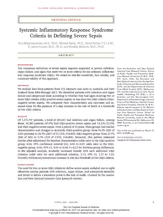
Patients presenting with AKI and multi-organ failure have been reported to have mortality rates of over 50%.
If CRRT required the mortality rate rise further to as high as 80%.
Slide3Classic renal indications for CRRTRapidly rising U/Es. Hyperkalaemia
unresponsive to medical management.
Severe metabolic acidosis.
Diuretic-resistant pulmonary oedema.
Oliguria
or
anuria
.
Slide4Non-renal indications for CRRT:Management of fluid balance e.g. cardiac failure.Clearance of ingested toxins.Correction of electrolyte abnormalities.
Temperature control.
Removal of inflammatory mediators in sepsis.
Slide5Implications of AKI:AKI follows from a rapid deterioration in renal function – especially in glomerular filtration rate (GFR) resulting from ischaemic or toxic injury to the kidney.
Loss of urine excretion –
anuria
. Raised levels of urea and
creatinine
; metabolic acidosis and electrolyte imbalances.
The inability to remove toxic metabolites is often associated with fluid overload – leading to pulmonary oedema and hypoxia as well as an increase in interstitial water.
May last days or weeks. Early correction is crucial.
Slide6Diagnosing of AKI:KDIGO guidelines define AKI as:Increase in serum Creatinine by ≥ 0.3mg/dl (≥ 26.5µmol/l) within 48 hours or
Increase in serum
Creatinine
to ≥ 1.5 times baseline, which is known or presumed to have occurred within the prior 7 days or
Urine volume ≤ 0.5ml/kg/hr for 6 hours.
Kidney Disease: Improving Global Outcomes (KDIGO) 2012
Slide7Staging of AKI:
Slide8Pathophysiology of AKI:The kidneys are particularly susceptible to damage as the renal parenchyma and the nephrons
are exposed to particularly high concentrations of toxins as water is
reabsorbed.
Additionally, there are regions in the kidney where oxygen supply is already
scarce under physiological conditions.
Any impairment of oxygen supply/perfusion could result in tissue damage.
Even though tubular necrosis may occur, regeneration of epithelial cells is
possible if the tubular basement membrane is damaged then the
nephron
will not
regain its function.
Slide9Aetiology:
Slide10Pre-renal: Results from decreased blood flow to the kidneys. There is no structural damageto the kidney in the early phase of pre-renal failure, but decreased oxygen supplycould result in tubular necrosis i.e. additional intrinsic renal failure.
Causes include
:
Dehydration due to poor fluid intake or excess fluid loss
Haemorrhage
Congestive heart failure
Sepsis
Poor systemic vascular tone
Poor renal vascular tone due to NSAIDS, ACE inhibitors or
hepatorenal
syndrome.
Slide11Intrinsic renal failure:Involves direct injury to the renal parenchyma and often includes the development of acute tubular necrosis (ATN).Causes include
:
Acute or rapidly progressive
glomerunephritis
Acute tubular necrosis caused by
ischaemia
or
nephrotoxic
drugs such as ACE inhibitors, acyclovir, high dose NSAIDs
Septic AKI mediated e.g. by inflammatory mediators or changes in intra-renal
haemodynamics
.Allergic or idiopathic interstitial nephritis Micro-vascular diseases such as
vasculitis
, disseminated intravascular coagulation (DIC), thrombotic thrombocytopenic
purpura
.
Slide12Post-renal failure:Results from obstruction of urine flow out of the kidney. Back-flow and backpressure result in increased intra-tubular pressure and a decreased filtration gradient.
Causes include:
Renal calculi
Tumours – these may be within the bladder or external to the renal system but apply pressure/obstruct outflow to the bladder,
ureter
or urethra.
Enlarged prostrate
Congenital malformations
Slide13Goals of treatment:Prevent further damage by treating the precipitating cause.Restore adequate cardiac output.Correction of electrolyte imbalance.
Correction of acid-base balance.
Removal of inflammatory mediators.
Prevention of fluid overload/access for nutritional support.
Slide14Overdose with a dialysable drug or toxin:Some drugs are removed by RRT but some are not.Drugs are cleared if they are water-soluble.
Drugs that are highly protein-bound are not cleared.
National Poisons Information Service
Slide15RRT techniques used in ICU:Treatment may be intermittent or continuous.Intermittent haemodialysis (IHD) or conventional renal dialysis involves renal unit nursing staff.
IHD method of solute clearance is by diffusion.
Continuous renal replacement therapy (CRRT) is used the most frequently involving ICU nursing staff.
CRRT method of solute clearance can is by convection (CVVH) or can be combined with diffusion (CVVHDF).
Slide16Filter anatomy:Thousands of fine capillaries.Blood flows through the fibres.Fluid surrounds the fibres.
Fibres are semi-permeable.
Similar to
glomerular
filtration.
Depending on the molecular weight,
various weight products can be filtered.
Slide17Solute clearance and molecules: Molecular weights up to 50,000 daltons are removed.The larger the molecule the slower the rate of transfer across the membrane.
E.G. urea (60) is cleared more efficiently than
creatinine
(113).
Small molecules <500 are cleared efficiently by diffusion but as molecule size increases diffusion becomes less effective
.
Middle molecules > 500 are cleared efficiently by convection
Slide18The smaller the molecule, the greater the clearance by
dialysate
/blood flow increases.
Slide19Slide20Drug prescribing:Drug adjustment is often required.Access to a pharmacist is advisable.Drug levels should be measured where possible.
Care is needed not to under-dose patients
especially
when using antibiotics to treat sepsis.
Refer to ‘The Renal Drug Handbook’ , online calculators or apps
Slide21Methods of clearance:ConvectionDiffusion
Slide22Convection:Relies on a pressure gradient.Solute removal is by solvent drag across the semi-permeable membrane.Convection extremely efficient at middle molecule clearance (>500
Da
).
Less efficient at small molecule clearance (<500
Da
)
but capable
of clearance – just takes longer.
Slide23Advantages of CVVH convection:Best suited for haemodynamically unstable patients.Creates less
haemodynamic
disturbance.
Gentler means of lowering urea and electrolytes.
Prevents large fluid shifts over short time.
Better control of fluid balance.
More flexibility for drugs and nutrition.
Clearance of molecules > 500 Da.
Slide24Disadvantages of CVVH (convection):Slower at small molecule clearance (<500 Da).Anticoagulation may be needed.
Heat loss.
Patient immobility.
More labour intensive and more expensive than IHD.
Slide25Dialysis (diffusion):Relies on a concentration gradient.Solutes move across membrane from an area of HIGH concentration to an area of
LOW
concentration.
High concentration = blood. Low concentration = replacement fluid.
Replacement fluid flows counter-current to direction of blood flow.
Replacement fluid (
dialysate
) is pumped from the green scale on the
prismaflex
.
Diffusion more efficient at small molecule clearance (<500
Da
).
Slide26Advantages of Renal Unit dialysis:Intermittent treatment.Short treatment ~ usually 3 to 4 hours ~ 3 times a week.Rapid removal and clearance of toxins (<500
Da
).
Rapid removal of fluid.
Slide27Disadvantages of Renal Unit dialysis:Poorly tolerated by haemodynamically unstable patients.Hypotension.
Arrhythmias.
Muscular complications.
Disequilibrium syndrome.
Slide28Slide29Slide30Pre Dilution:
Slide31Benefits of Pre Dilution:Dilutes blood before it comes in to contact with the hollow fibres.Dilutes the blood in the filter fibres.Reduces concentration of clotting factors.
Prolongs the life of the filter.
Slide32Post Dilution:
Slide33Benefits of Post Dilution:Enhanced clearance of toxins > 15%.Speedier clearance of potassium; urea; creatinine; acidosis and inflammatory mediators.
Possible earlier reduction in
vasoactives
.
Possible reduced length of treatment time.
Possible earlier enhanced recovery and earlier discharge from ICU.
Slide34Vas-cath or dual-lumen catheter:Right internal jugular or right subclavian ~ a 15cm length catheter may be used
Left internal jugular or left
subclavian
~ a 20cm catheter may be inserted the full length or to the black guide mark.
Femoral access: 25cm length may be used and inserted to the black guide mark. Bio-patch is used for femoral sites.
Slide35Insertion Procedure:As per CVC insertion.Trendelenburg.CVC insertion sticker needs to be completed and filed in medical notes.
Vascath
maintenance sheet needs to be completed.
Slide36When to change the filter:End of filter life i.e. at 72 hours.If blood appears in the waste collection bag (turns pink) → triggers the ‘blood leak detected’ alarm.Blood in waste bag indicates filter leak/rupture and treatment needs to be terminated immediately
.
If filter leak/rupture occurs – complete
datix
form and inform Gambro
If BLD alarm occurs – check that the front panel of the machine is not in direct sunlight
Slide37Catheter patency:20ml sodium chloride 0.9% via each port.Heparin lock (1000 units per ml vial) → Lock with volume indicated on the catheter ports
→
Use red caps.
Always remove the
same volume
from
vascath
prior to recommencing treatment.
This ensures residual heparin is removed from the dead space.
Slide38Slide39Slide40Anticoagulation via the haemofiltration circuit: 1. Citrate
2. Heparin
“For anticoagulation in CRRT, we suggest using regional citrate
anticoagulation rather than heparin in patients who do not have
contraindications for citrate” KDIGO guidelines 2012
Slide41Slide42Coagulation cascade
Slide43Citrate – is metabolised in liver, skeletal muscle and the kidney into bicarbonate releasing the chelated calcium
Slide44Slide45Approximately 50% calcium is lost in the effluent
Slide46Citrate dose to prevent clotting:Concentration of citrate required to inhibit extracorporeal coagulation has been estimated at approximately between 2.5 and 5.0 mmol/L blood in pharmacodynamic studies.
No clotting factors if ionised calcium level of the extracorporeal circuit is maintained between 0.25 and 0.35
mmol
/L.
Prismaflex
administers citrate solution into the circuit at a dose to maintain circuit ionised calcium levels between 0.25 and 0.35mmol/L
post filter
– default starting dose is 3.00mmol/L blood.
Slide47Net loss of calcium: blood returning to the patient combines with venous blood in body normalising the ionised calcium and preventing systemic anticoagulation but there is a net loss of calcium-citrate complex into the effluent.
Slide48Slide49The prismaflex syringe driver compensates for the net loss:Citrate anticoagulation is only regional – returning extracorporeal blood mixes with systemic blood; calcium (therefore clotting) normalises instantly.
However, there is a loss of calcium-citrate complex into effluent through convection and/or diffusion.
Calcium needs to be infused into the patient to replace the loss to the effluent.
The syringe pump automatically delivers calcium to the patient at a dose to replace calcium lost into the effluent – default 100%. The rate will adjust automatically when other flow rates are altered to maintain percentage.
Slide50Solutions for use during Citrate Anticoagulation in CRRT
Doesn’t contain K+ 4mmol/litre K+ 4mmol/litre K+
Slide51Slide52Equipment needed:ST150 Prismaflex® kit CA250 calcium line
50 ml Luer lock syringe
5 litre bag of
Prismocitrate
® 18/0 (citrate used as pre-dilution)
5 litre bag of Prism0cal® B22 (calcium-free
dialysate
solution)
5 litre bag of Prismasol4® (post-dilution replacement solution)
Slide53Equipment needed:2 x 1 litre bags of 0.9% sodium chloride (priming solution) 3 ampoules of calcium chloride diluted to total volume of 50ml using 0.9% sodium chloride. Sterile dressing pack and trolley
2 x 2 ml syringes to withdraw hep-lock, 2 x 20 ml syringes to check flow (20 ml in 6 seconds), 2 x 10 ml syringes for sodium chloride flush.
Slide54Setting up and Priming:Switch the machine on, input the patient details, ensure accurate patient weight and haematocrit is entered. Choose the CVVHDF option
→
Choose citrate-calcium via the
Prismaflex
®
syringe pump.
Follow the on-screen step-by-step installation instructions.
Install
Prismocitrate
® 18/0 on the white scale (PBP = pre-blood pump).
Install Prism0cal® B22 on the green scale (
dialysate).Install
Prismasol
4 on the purple scale (replacement)
Slide55Setting up and priming:Prime the ST150 with 2 litres of 0.9% sodium chloride (no heparin required).Install the calcium chloride syringe into the Prismaflex® syringe pump. This should be a 50ml
Luer
Lock syringe.
Leave the calcium line unclamped for priming by the
Prismaflex
® machine.
Ensure fluid loss/gain limit is set to 400 ml/3 hours, this is a default setting do not change.
Slide56Starting parameters:Mode: CVVHDF Starting citrate dose: 3.0 mmol/L blood
Starting calcium compensation: 100%
Flow settings: Based on actual body weight
Slide57Connection:Using a sterile pack and sterile gloves to access the vascath, ensure flow test isperformed –
1. Withdraw 5 ml blood from the red
vascath
port to remove hep-lock and discard onto gauze (observe for clots).
2. Withdraw and replace 20 ml blood. Flow should be sufficient to withdraw 20 ml blood from the
vascath
in 6 seconds or less.
3. Flush with 10 ml 0.9% sodium chloride.
4. Repeat on the blue
vascath
port.
Slide58Monitoring example:Measure serum electrolytes and ABGS before initiation.At one hour
measure
(1) post filter ionised calcium to check circuit calcium
(2) patient ionised calcium to check systemic calcium
Adjust citrate dose or calcium concentration according to table.
Make adjustments as required and repeat measurement after
one
hour.
Once ideal values/steady state reached measure 6 hourly
Once a day total calcium to check ratio.
Slide59Example monitoring table – clinical decision support
Slide60Treatment adjustments:
Slide61Slide62Slide63Slide64