
Pelvis Models Rudiger Anatomia and 3B Scientific https3d4medical8K6xxapi Figure 12 Figure 11 Rudiger Anatomie Model Rectal Hiatus Anal Aperture Urogenital Hiatus Urogenital Lab 1 Station 2 ID: 934115
Download Presentation The PPT/PDF document "Urogenital Lab 2, Station 2" is the property of its rightful owner. Permission is granted to download and print the materials on this web site for personal, non-commercial use only, and to display it on your personal computer provided you do not modify the materials and that you retain all copyright notices contained in the materials. By downloading content from our website, you accept the terms of this agreement.


Slide1
Urogenital Lab 2, Station 2
Pelvis Models:
Rudiger
Anatomia
and 3B Scientific
Slide2https://3d4medic.al/8K6xxapi
Figure 1.2
Figure 1.1:
Rudiger
Anatomie
Model
Rectal Hiatus (Anal Aperture)
Urogenital Hiatus
Urogenital, Lab 1: Station 2
Levator
Ani
Obturator
Internus m.
Iliococcygeus m.
Pubococcygeus m.
Puborectalis
m.
Coccygeus
Piriformis
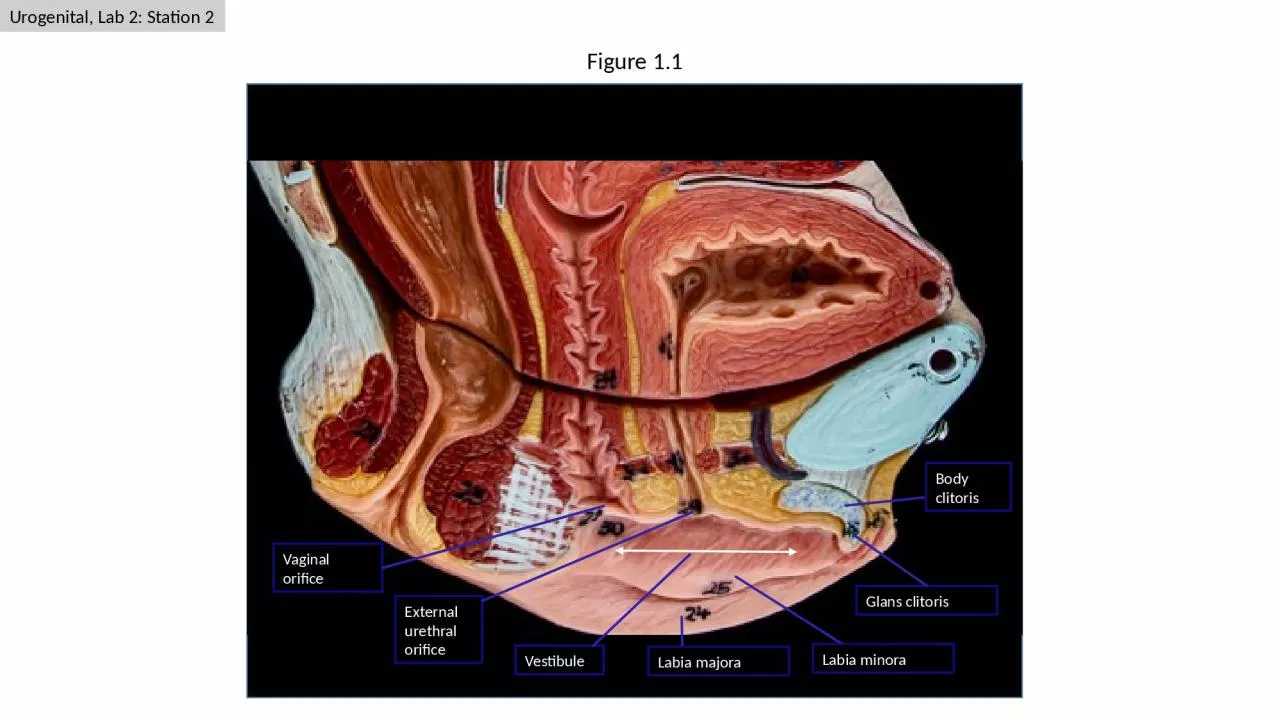
Bladder
Uterus
Rectum
Vagina
Tendinous arch
Greater Sciatic
Foramen
Slide3Obturator internus m.
Piriformis m.
Coccygeus
m.
Levator
ani
Tendinous arch
Figure 1.3:
Figure 2.1
Levator
ani
Obturator internus m.
Figure 1.4
3B Scientific Model
Figure 1.3
3B Scientific Model
Urogenital, Lab 1: Station 2
Slide4rectal hiatus
(anal aperture)
Figure 2.1
Figure 2.1
Urogenital triangle
Anal triangle
Figure 2.2
Figure 2.3
Superficial transverse perineal m.
Urogenital, Lab 1: Station 2
Sacrotuberous ligament
Coccyx
Ischiopubic
ramus
Slide5Figure 2.4
Figure 2.5
Figure 2.1
Ischioanal
fossa
Anal canal / Anus
External anal sphincter
Inferior rectal n.
Pudendal n.
Urogenital, Lab 1: Station 2
Slide6Perineal Membrane
Figure 3.2
Figure 3.1
Bulbospongiosus m.
Perineal body
Superficial transverse perineal m.
Greater vestibular (Bartholin) gland
Bulb of Vestibule
Urogenital, Lab 1: Station 2
Figure 3.3
Glans clitoris
Perineal Membrane
(green tape)
Ischiocavernosus m.
Slide7Pelvic Diaphragm
Superficial Pouch
Deep Pouch (Fibromuscular Region)
“Urogenital Diaphragm”
Deep Pouch (A Recess
Ischioanal
Fossa)
Perineal Membrane
Colles
Fascia
Figure 3.4
Figure 3.6
Figure 3.5
Perineal Membrane
(green string)
Muscle in deep pouch
(“fibromuscular” region or formerly named urogenital diaphragm)
Urogenital, Lab 1: Station 2
Urethra
Deep perineal space (pouch)
Anterior recess of ischioanal fossa
Glans clitoris
Body clitoris
Perineal body
Slide8Sacral ventral
rami (S1-S4)
Lumbosacral trunk
Lumbosacral plexus
Figure 4.1
Pudendal n.
Muscle in deep pouch
(deep to removed perineal membrane)
Anterior recess of ischioanal fossa
Perineal n.
Dorsal n. clitoris
Pudendal n.
Figure 4.2