
DISEASES How Proteins fold and why they misfold Role of Molecular Chaperones in Protein Folding ORGANELLESPECIFIC PROTEIN QUALITY CONTROL SYSTEMS AND PROTEIN MISFOLDING DISEASES Mechanisms and Link to Disease ID: 932824
Download Presentation The PPT/PDF document "PROTEIN MISFOLDING AND HUMAN" is the property of its rightful owner. Permission is granted to download and print the materials on this web site for personal, non-commercial use only, and to display it on your personal computer provided you do not modify the materials and that you retain all copyright notices contained in the materials. By downloading content from our website, you accept the terms of this agreement.


Slide1
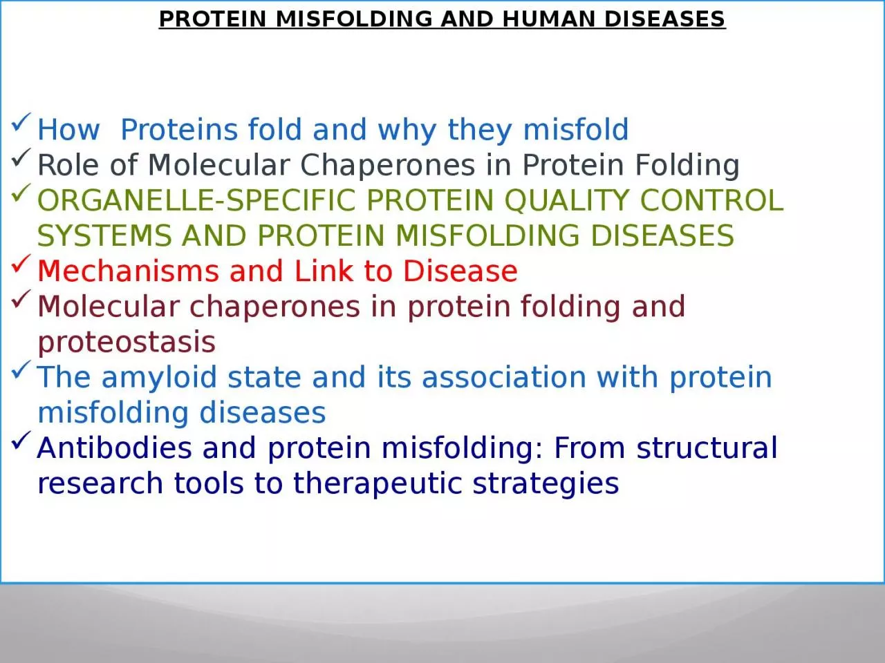
PROTEIN MISFOLDING AND HUMAN
DISEASES
How
Proteins
fold and why they
misfold
Role of Molecular Chaperones in Protein Folding
ORGANELLE-SPECIFIC PROTEIN QUALITY CONTROL SYSTEMS AND PROTEIN MISFOLDING DISEASES
Mechanisms and Link to Disease
Molecular chaperones in protein folding and
proteostasis
The amyloid state and its association with protein
misfolding
diseases
Antibodies and protein
misfolding
: From structural research tools to therapeutic strategies
Slide2PROTEIN MISFOLDING DISEASE: GAIN-OF-FUNCTION AND LOSS-OF-FUNCTION DISEASES
Aggregation of Copper–Zinc Superoxide Dismutase in Familial ALS
Protein
Misfolding
in Alzheimer Disease: The Aβ oligomer hypothesis for synapse failure and memory loss in Alzheimer’s disease
Mechanisms of emphysema in α1-antitrypsin deficiency: molecular and cellular insights
Protein
Misfolding
and Aggregation in Cataract Disease
Techniques for Monitoring Protein
Misfolding
and Aggregation in Vitro and in Living Cells
Identifying and validating biomarkers for Alzheimer’s disease
Slide3Protein folding and
misfolding
Slide4INTRODUCTION
The function of most cellular proteins is
dependent on
their
three-dimensional structure
, which
is acquired through folding
of the
polypeptide chain coded from the
nuclear genome.
Changes in
the polypeptide chain, either
resulting from
inherited or acquired gene variations
or from
abnormal
amino acid modifications
,
may change
the folding process and give rise
to
misfolding
of the protein.
Slide5Depending on the
nature
of the protein itself, the cellular
compartment
in which the
misfolding
occurs, the activity of the folding and degradation machineries as well as interacting genetic factors and the cell and environmental conditions, the
consequences
of the
misfolding
may
be quite
different
Slide6However, despite this diversity
, a
large number of
very different diseases
, from
early-onset inborn errors of
metabolism to
late-onset neurodegenerative diseases,
can be
viewed as
protein
misfolding
diseases
,
often called
conformational
diseases
.
On
this basis, a common framework
related to
the molecular pathogenesis and cell
pathologic mechanisms
in these very different
diseases is
emerging.
Slide7Aims of these lessons
W
e
introduce the theory
of
protein
folding and
misfolding
in general
and mention
the emerging ways to predict
consequences of
amino acid
alterations
We
introduce the
concept of
protein quality
control (
PQC)
and discuss the various
compartment specific PQC
systems as well as the
molecular pathogenesis
of some representative
misfolding
diseases
originating in the various compartments.
Slide8General facts on protein folding
Important elements for protein folding:
The amino acid sequence
The right cellular environment
The perfect balance between the various folding states
A fully functional Protein Quality Control (PQC) system
(T ,P ,pH ,
etc…
)
(Chaperones, proteasome unit )
Slide9Causes of
misfolding
and protein aggregation
How protein aggregates
form?
Change in cellular conditions
more
misfolded
proteins
the PQC system is overwhelmed
aggregation is favored
Aggregation is thought to be set in by protein segments containing hydrophobic amino acids residues, β
-sheet predisposition and low net charge.
Slide10Causes of
misfolding
and protein aggregation
States accessible to a protein molecule
Free-energy folding landscape for chaperone-mediated protein folding
Slide11Causes of
misfolding
and protein aggregation
Protein aggregation is a 2-stage event
The
nucleation
proteins start attaching reversibly to a growing nucleus
Proteins
attach irreversibly
to the nucleus until it becomes a larger aggregate
.
Slide12Cellular consequences of protein aggregation
Loss-of-function pathogenesis
:
if
misfolded
proteins are prematurely degraded by PQC system
protein deficiency disease
Gain-of-function pathogenesis
:
if
misfolded
proteins are not eliminated but accumulated instead
disease pathology toxicity
Some diseases display both pathogenic mechanisms.
Slide13Cellular consequences of protein aggregation
the slowing down of polypeptides translation
+
Slide14Protein-
misfolding
diseases
Include
conditions where a
protein:
fails to fold correctly
(cystic fibrosis,
Marfan
syndrome,
amyotrophic
lateral sclerosis)
is not stable enough to perform its normal function (many forms of cancer)
fails to be correctly trafficked (familial hypercholesterolemia, α1-antitrypsin deficiency)forms insoluble aggregates that deposit toxically (neurodegenerative diseases: Alzheimer’s, type II diabetes, Parkinson
’
s and many more)
Slide15The fundamental mechanism of protein folding
The concept of an energy landscape
Slide16Protein Folding
Slide17Slide18Slide19Slide20Slide21Slide22Protein folding models
As Anfinsen showed
the
amino
acid sequence
of a polypeptide chain contains
all necessary
information determining the
three-dimensional functional
structure of a
given protein.
This
means that the native state has to be
thermodynamically stable and the protein must rapidly find the native state. If a protein searches through all possible conformations in a random fashion until it finds the conformation
with the lowest free energy it will take an enormous amount of time.
Slide23Imagine a polypeptide chain with
100 residues
and every residue has
2
possible
conformations
. The protein has 2
100
(or 10
30
) possible conformations, and if it converts one conformation into another in the shortest possible time (maybe 10
-11
s) the time required is
1011 years. A protein however reaches its native fold in 10-3 to 103 s both in vitro and in vivo. The
Levinthal paradox states that a random search for the final conformation would take millions of years
Slide24To overcome this obstacle, nature uses a number of biochemical “rules” and has evolved some assisting components facilitating the folding processes, the so-called
molecular chaperones
(see later)
Slide25Molecular chaperones
assist the folding by protecting
the protein
during the folding process and
keeping it
away from
misfolding
, which may
lead to
aggregation
(see later).
The
target
for chaperones is unfolded and partially folded polypeptide chains with exposed stretches of hydrophobic amino acids that are usually inside in the core of folded proteins. Proteins with exposed hydrophobic
stretches are reversibly bound to and released from the chaperones. Many chaperones have an ATPase domain that orchestrates conformational changes,
switching the molecule between high and low binding affinity states
Slide26Slide27ϕ
and
ψangles
The planarity of the peptide bond means that there are only two degrees of
freedom per
residue for the peptide chain. Rotation is allowed about the bond
linking the α-carbon
and the carbon of the peptide bond and also about the bond linking the nitrogen of the peptide bond and the adjacent
α-
carbon
.
The angle
about the
Cα-N bond is denoted by the Greek letter ϕ (phi), and that about the Cα-C is denoted by ψ (psi).
Slide28The driving force of protein folding is
the search
for a conformation with
lower
free
energy than
the previous one.
The various
lower energy
states can be separated by barriers
of higher
energy, and to overcome these
barriers chaperone
assistance may be required.Biophysical measurements
and computer simulations have revealed that many of the local elements of protein structures can be generated very rapidly; for example, individual
α-helices are able to form in less than 100 ns, and β-turns in as little as 1 μs . Indeed, the folding in vitro
of some of the simplest proteins,
is
completed in less than 50
μs
Slide29A certain
hierarchy of interactions
seems to exist between residues for the first part of the folding process, the so-called
nucleation- condensation process
, which speeds up the folding through a number of
transition states
characterized by the presence of interatomic interactions also present in the native protein
Slide30To picture protein
folding it has
been
suggested
a
topological landscape
representing different energy
levels
In this model the
process of
folding is described as a constant
striving toward
minimal free energy with the
native structure of the protein being the conformation with
the lowest energy level, the global minimum of the landscape
Slide31The
landscape is
drawn as a
funnel
shape
in a
three-dimensional system with the
free
energy on
the y axis
and the
conformational space
or
entropy as a two-dimensional
projection on the x and z axes.
Slide32Because the free energy of a protein is
a function
of its conformation defined by
the
interactions
between the amino acid residues
, even
small alterations
in the amino acid
chain may
change the surface of the landscape,
leading to
the possible formation of
new local free energy minima resulting in a different stable structure of the protein, which may be prone to aggregation.
Slide33To
refine the
concept of energy folding
landscapes to
include the aggregation tendency,
which may
be
aggravated by extrinsic factors
, such
as
high
protein concentration
and
elevated temperature, in the living cell, one can imagine a landscape with two deep valleys
Slide34Free-energy folding landscape for chaperone-mediated protein folding
:
hypothetical landscape of all possible protein conformations pictured with
higher altitude
symbolizing
higher free energy and entropy.
Chaperones
iteratively bind and release their substrates, each time raising
the free energy and enabling escapes from wrong folding pathways
, indicated
by the “
Unfolding
” arrow. Eliminating protein by degradation occurs mainly in areas with trapped misfolding
Slide35Energy landscapes can
have
many
different shapes
and have
many “hills”
which represents the
high
energy conformations
that sometimes has to be passed to reach the native state.
Protein aggregates
can be extremely stable, even more stable than the native protein, suggesting that the aggregated states are trapped in the kinetically deepest valleys of the landscape.
Slide36PROTEIN QUALITY CONTROL
SYSTEMS
In the
test tube
, protein folding is
performed most
efficiently at
low protein
concentrations and
low temperature.
In
contrast,
protein folding
in human cells takes place at 37◦C—or higher during fever—with very high concentrations
of proteins. To sustain a reasonable efficiency of protein folding in this challenging environment and to protect againstthe negative consequences of environmental changes
, PQC systems have evolved that supervise folding, counteract aggregation, and eliminate misfolded and damaged polypeptide chains before they can exert toxic effects.
Slide37Quality control mechanism
Regulation of protein folding in the ER
.
Many
newly synthesized
proteins are
translocated
into the ER
, where they fold into their three-dimensional structures
with the help of a series of molecular chaperones and folding catalysts (not shown).
Correctly
folded
proteins
are then transported to the Golgi complex and then delivered to the extracellular environment. However, incorrectly folded proteins
are detected by a quality-control mechanism and sent along another pathway (the unfolded protein response) in which they are ubiquitinated
and then degraded in the cytoplasm by proteasomes
Slide38Important components of
these systems are comprised by
molecular chaperones
.
Additionally
, the PQC
systems contain
specialized
intracellular proteases
and
accessory
factors
that regulate the activity
of chaperones and proteases or provide communication between the various components.
Slide39Although there is some
overlap and
certain chaperones are able to
fulfill several
functions, one can distinguish between
folding chaperones
,
holding chaperones
,
and
unfolding
chaperones
.
Folding chaperones promote folding, whereas holding chaperones, like the small heat-shock proteins lacking an ATPase activity, reversibly bind misfolded proteins. They need to transfer their substrates to folding chaperones to
accomplish folding. Unfolding chaperones typically contain one or two so-called AAA+
domains that harbor an ATPase, and are able to unfold misfolded proteins, either for degradation or to give them a new start for folding
Folding, holding and unfolding chaperones
Slide40Proteases
of PQC systems
The proteases of PQC systems have a
particular structure
secluding the
proteolytically
active
sites
inside cavities that are not
accessible for
folded proteins
To be degraded, proteins
must first be
fully unfolded before they are injected into the proteolytic
cavities, where they are processively degraded into small peptides. This is typically accomplished by AAA+ domain containing unfolding
chaperones.
Slide41Functioning of protein quality control (PQC) systems.
PQC
systems
manage the pool of unfolded
and partially
folded conformations
(
center
).
Folding
chaperones
promote folding,
holding
chaperones maintain solubility, and unfolding chaperones disaggregate aggregates or unfold misfolded proteins andinject them into proteolytic chambers of PQC proteases
Slide42The
compartmentation
of the
cellular PQC systems