
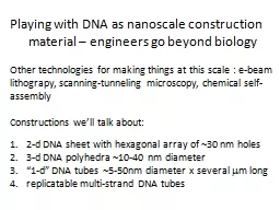
ENT Procedures Operative Sequence Myringotomy Define the procedure A small incision is made in the tympanic membrane to allow the drainage of fluid from the middle ear and the placement of ear tubes ID: 931584
Download Presentation The PPT/PDF document "Myringotomy with Ear Tubes" is the property of its rightful owner. Permission is granted to download and print the materials on this web site for personal, non-commercial use only, and to display it on your personal computer provided you do not modify the materials and that you retain all copyright notices contained in the materials. By downloading content from our website, you accept the terms of this agreement.


Slide1
Myringotomy with Ear Tubes
ENT Procedures
Operative Sequence
Slide2MyringotomyDefine the procedure:A small incision is made in the tympanic membrane to allow the drainage of fluid from the middle ear and the placement of ear tubes.
Slide3MyringotomyOverall Purpose of Procedure:Relieve effusion.Effusion = ‘s fluid in the middle ear causing painful ear infections (a.k.a. acute otitis media)
Acute otitis media - Inflammation of the middle ear in which there is fluid in the middle ear accompanied by signs or symptoms of ear infection: a bulging eardrum usually accompanied by pain; or a perforated eardrum, often with drainage of purulent material (pus).
Slide4Slide5Ear TubesEar tubes are tiny cylinders placed through the ear drum (tympanic membrane) to allow air into the middle ear. They also may be called tympanostomy tubes, myringotomy tubes, ventilation tubes, or PE (pressure equalization) tubes. These tubes can be made out of plastic, metal, or Teflon and may have a coating intended to reduce the possibility of infection.
There are two basic types of ear tubes: short-term and long-term.
Slide6Ear Tubes cont.
Short-term tubes are smaller and typically stay in place for six months to a year before falling out on their own. Long-term tubes are larger and have flanges that secure them in place for a longer period of time. Long term tubes may fall out on their own, but removal by an ENT surgeon is often necessary.
Slide7MyringotomyWound Classification: 2
Slide8Operative Sequence1- Incision2- Hemostasis3- Dissection
4- Exposure5- Procedure (Specimen Collection possible)6- Hemostasis 7- Irrigation 8- Closure
9- Dressing Application
Slide9MyringotomyInstrumentation: Myringotomy tray, microscope, speculum holder, micro-instruments.
Positioning: The patient is in supine position arms tucked. MD will sit for procedure.Prepping: Surgeon preference. Hibiclense or a Betadine Prep Kit. Betadine can be placed on cotton ball to swab the ear if MD prefers. Some use no prep at all...always ask
Draping
: 4 towels and a head drape. Ask about towel clips. Ask about clear incise drape.
Slide10Myringotomy Begin your Operative SequenceIncision: made into the tympanic membrane with
myringotomy blade (this step is out of order in this surgery due to the fact that we already have an opening to work through – the ear canal)
Slide11Myringotomy cont. Operative SequenceHemostasis: noneDissection and Exposure
: ear spec and microscopeExploration and Isolation: MD will remove ear wax (cerumen) with ear curettes and visualize the TM.
Slide12Myringotomy cont. Operative SequenceSurgical Repair/Removal/Specimen Collection:
Will make a 2mm to 3mm incision into the TM.Fluid is suctioned out with micro-Frasier suction, #3 or #5.
Slide13Myringotomy cont. Operative SequenceSurgical Repair/Removal/Specimen Collection
:PE tube is grasped either by the MD or scrub with alligator forceps.Placed into the incision.Rosen needle (actually a pick) used to manipulate PE tube into correct position
.
Video
http://www.youtube.com/watch?v=j_Z-ylTtRts
Slide14Myringotomy cont. Operative SequenceHemostasis and Irrigation:noneClosure:
Places antibiotic drops into ear canal. Places cotton ball in ear canal.
Slide15MyringotomyMajor Arteries:Posterior auricular artery
Labyrinthine artery Major Veins:Posterior auricular vein
Slide16MyringotomyMajor Nerves: Vestibular nerve: The vestibular nerve is one of the two branches of the
Vestibulocochlear nerve (the cochlear nerve being the other). It goes to the semicircular canals via the vestibular ganglion. It receives positional information.
Slide17Tonsillectomy
& Adenoidectomy
ENT Procedures
Operative Sequence
Slide18Tonsillectomy and AdenoidectomyTonsils are small, round pieces of tissue that are located in the back of the mouth on the side of the throat. Tonsils are thought to help fight infections by producing antibodies.
Slide19Adenoid - Lymph-like areas of tissue, or glands, that are similar to the tonsils, but they are located very high in the throat, behind the nose. They trap and filter out germs that enter the body. The adenoids also help your body fight off infection by making antibodies.
Slide20Types of TonsilsPalatine tonsils--located on each side of the throat.
Pharyngeal tonsils--also known as adenoids are near the posterior openings of the nasal cavity.
Lingual tonsils
--near the base of the tongue
Slide21Slide22You have your adenoids when you are born and they continue to grow until you are 5 to 7 years old. By school age, the adenoids begin to shrink in size, and, by the time children reach their pre-teen or teenage years, the adenoids are usually small enough to not cause any symptoms.
Slide23What Are the Symptoms of Enlarged AdenoidsA child may complain of:difficulty breathing through the nose is breathing through the mouth
talks as if his or her nostrils are pinched breathes noisily snores while sleeping stops breathing for a few seconds while sleeping (called sleep apnea)
Slide24Tonsillectomy and AdenoidectomyDefine the procedure: removal of tissue to eradicate infection, improve the airway or remove cancer.
Slide25Tonsillectomy and AdenoidectomyOverall Purpose of Procedure:3 pathological indications for removal of the tonsils and
adnoids:InfectionHypertrophy- enlargement via cellular growth
Cancer
Pt. may suffer from tonsillitis,
peritonsillar
abscess, strep throat,
irr
. sleep patterns, difficulty swallowing.
Adenoids can become hypertrophic to the point of blocking the Eustachian tube, causing
otitis
media.
Slide26Slide27Tonsillectomy and AdenoidectomyWound Classification: 2
Slide28Operative Sequence1- Incision2- Hemostasis
3- Dissection 4- Exposure5- Procedure (Specimen Collection possible)
6- Hemostasis
7- Irrigation
8- Closure
9- Dressing Application
Slide29Tonsillectomy and AdenoidectomyInstrumentation: T&A tray
Positioning: The patient is in supine position arms tucked. MD will sit for procedure. Spin bed 90 degrees for some Md’s.
Prepping
: NONE!
Draping
: 4 towels and a head drape (depends on MD). Ask about towel clips. Down sheet.
Slide30Tonsillectomy and Adenoidectomy Begin your Operative Sequence
Incision: This step comes later in the procedure.Hemostasis: none at this point in the procedure.
Slide31Tonsillectomy and Adenoidectomy cont. Operative Sequence
Dissection and Exposure: Mouth Gag of Md choice is placed into pt’s mouth. Mouth gag WILL REST on mayo stand.
Slide32Tonsillectomy and Adenoidectomy cont. Operative SequenceExploration and Isolation:
MD might have headlight for visualization purposes.Tonsil is grasped with Allis clamp, possible Allis Adair.Have FRED on mayo for dental mirror.
Slide33Tonsillectomy and Adenoidectomy cont. Operative Sequence
Surgical Repair/Removal/Specimen Collection:An incision is made in the grasped tonsil.Incision can be made with a Snare, Laser, Curette, or Coblation Wand.
Tonsil is removed with the device of choice.
Scrub needs to have a chromic suture ready on the table for heavy bleeding.
Slide34Tonsillectomy and Adenoidectomy cont. Operative Sequence
Surgical Repair/Removal/Specimen Collection:Coblation wand uses radiofrequency waves, instead of
cautery
(heat) techniques, to remove tonsils and adenoids.
Coblation
Tonsillectomy:
http://www.youtube.com/watch?v=KizSZuqkyBc
Slide35Tonsillectomy and Adenoidectomy cont. Operative SequenceSurgical Repair/Removal/Specimen Collection
:You will repeat the same process for the other tonsil.Adenoid – MD will retract the palate with a red rubber catheter inserted
transnasally
.
Clamp end of red rubber with Kelly.
MD will use dental mirror to view adenoid tissue.
Removal with same procedure as Tonsils.
You will have specimens – ask then pass off.
May need sterile safety pin to mark specimen. (usually the safety pin will go into the right tonsil. Be sure not to put the pin into your hand – Tonsils are very dirty)
Slide36Tonsillectomy and Adenoidectomy cont. Operative Sequence
Hemostasis and Irrigation:Heavy bleeding is a possibility. Always have NACL ready on back table or mayo.
Closure
: packed with strung gauze. May be soaked in viscous
Lidocaine
.
T and A
vid
Child and Adult T&A EESEDU
NEVER BREAK YOUR TABLE DOWN
Slide37Tonsillectomy and AdenoidectomyMajor Arteries:
tonsillar and ascending palatine branches of the facial artery
Major Veins:
tonsillar veins
Slide38Tonsillectomy and Adenoidectomy
Major Nerves: tonsillar branches of glossopharyngeal nerve
Slide39SUR 122Tracheotomy/Tracheostomy
Slide40Tracheostomy is indicated for a patient who requires emergent or elective airway management for:prolonged ventilator dependence acute upper airway obstructionchronic upper airway obstruction
Slide41Pathology for Tracheotomy or TracheostomyVocal cord paralysisNeck surgeryTrauma
Prolonged intubationSecretion managementCannot intubateStridor due to tracheal blockageSleep apnea
Slide42Anatomy of the Neck
(From Potter PA and Perry AG: Fundamentals of nursing, ed 5, St Louis, 2001, Mosby.)
Slide43Anatomy of the LarynxAnterior view of the pharynx
Posterior view
of the pharynx
(From Thibodeau GA and Patton KT: Anthony's textbook of anatomy and physiology, ed 17, St Louis, 2003, Mosby.)
Slide44Tracheotomy/TracheostomyTracheotomy temporary opening into the trachea to facilitate breathing Tracheostomy permanent opening of the trachea and creation of a tracheal stoma
Must place tracheal tube with eitherPatient will be hooked up to a ventilatorLong term tracheostomy may eventually be able to wean off ventilator, but maintain stoma that will function as their nose did prior to surgery
Slide45AnesthesiaGeneralLocal
Slide46MedicationsLocal anesthetic: Lidocaine or bupivicaine with or without epinephrineAntibiotic irrigation
Slide47PositioningSupineShoulder rollDonut headrestPillow under kneesSafety strap
Slide48Prep End of chin to midchest and bedsheet to bedsheetPrep of choice: Duraprep, betadine scrub and/or paint
Slide49DrapingTowels Small fenestrated sheet (Pediatric lap sheet)
Slide50Supplies, Equipment, InstrumentsMinor basinBasic packPediatric lap sheetOther small fenestrated sheet
Blades Suture or ties of surgeon’s choice (prn)Tracheotomy tray
Tracheotomy tube (Shiley)
Twill tape
Slide51Operative SequenceDiscussion
Slide52Surgical ProceduresTracheotomy/Tracheostomy
Isthmus of thyroid is divided to expose
the trachea
Tube is inserted
Two tracheal rings are cut, and the upper ring is partially resected. Tracheal hook pulls the trachea from the depth of the wound toward the surface
(Modified from DeWeese DD: Textbook of otolaryngology, ed 6, St Louis, 1982, Mosby.)
Slide53ConsiderationsWill make sure obturator goes with patient to PACU or ICUComplications: hemorrhage, infection, damage to other structures
TRACHEOSTOMY Video
Slide54ENT Procedures
Operative Sequence
Septoplasty
Slide55Slide56Septum anatomyThe nasal septum separates the left and right nasal airway. The yellow portion is made of flexible cartilage, the quadrangular cartilage. The blue portion is thin bone, the perpendicular plate of the ethmoid bone. The purple portion is thicker bone, the vomer bone.
Slide57Slide58SeptoplastyOverall Purpose of Procedure:To produce a patent nasal airway
Slide59SeptoplastyDefine the procedure:
A surgical procedure done to improve the flow of air to your nose by repairing malformed cartilage and/or the bony portion.
Slide60Indications 1. Mouth breathing,
2· Snoring, 3· Drooling during sleep, 4· Change in voice,
5· Decrease sense of smell and taste
6· Sometimes sleep disturbances.
7· The symptoms are usually worse on one side, and sometimes occur on the side opposite the bend.
In some cases the crooked septum can interfere with the drainage of the sinuses, resulting in repeated sinus infections.
The septum may also need to be straightened in individuals undergoing sinus surgery just so that the instruments needed for this operation can be fit into the nasal cavity.
Slide61SeptoplastyThis procedure may be performed in conjunction with:Rhinoplasty
–a facial cosmetic procedure, usually performed to enhance the appearance of the nose. During rhinoplasty, the nasal cartilages and bones are modified, or tissue is added. The aim is to improve the visual appeal of the nose. Rhinoplasty is also frequently performed to repair nasal fractures. When rhinoplasty is used to repair nasal fractures, the goal is to restore pre-injury appearance of the nose.
Sinus surgery
Slide62SeptoplastySeptoplasty is rarely performed in children because the septum is is
the major growth center of the midface; disrupting it may lead to maxillary hyperplasia (enlarged upper jaw)Wound Classification
: 2
Slide63Operative Sequence1- Incision2- Hemostasis
3- Dissection 4- Exposure5- Procedure (Specimen Collection possible)
6- Hemostasis
7- Irrigation
8- Closure
9- Dressing Application
Slide64SeptoplastyInstrumentation: Septoplasty tray
Positioning: The patient is in supine position arms tucked. MD will sit for procedure. Spin bed 90 degrees for some MD’s.
Prepping
: NONE!
Draping
: 4 towels and a head drape (depends on MD). Ask about towel clips. Down sheet.
Slide65Septoplasty Begin your Operative Sequence
Incision: before this step comes the MD will inject the nose and turbinates with 1% Lidocaine with Epi. Then MD will pack the nose nasal packing soaked in vasoconstrictor of choice. (Afrin, Cocaine, Adrenaline)Hemostasis: epi
Slide66Septoplasty cont. Operative Sequence
Dissection and Exposure: Placement of nasal speculum
Slide67Septoplasty cont. Operative SequenceExploration and Isolation:
MD might have headlight for visualization purposes.May perform with the aid of a scope
Slide68Septoplasty cont. Operative Sequence
Surgical Repair/Removal/Specimen Collection:Incision is made into the septum below the obstruction.Small Tenotomy
scissors are used to dissect the membranous nasal septum and expose the cartilage.
Slide69Septoplasty cont. Operative Sequence
Surgical Repair:A Freer or Cottle elevator is then used to elevate the septum off the underlying tissue.MD removes the deviated bone using a small chisel and mallet.
Pieces are then removed with a Takahashi forcep
Incision repair with 4-0 Chromic suture
Slide70Septoplasty cont. Operative Sequence
Hemostasis and Irrigation:NACLSuction-
Bovie
if needed.
Closure
: internal nasal splints are placed
bilat
. to stabilize the septum and are stitched to the membranous septum with a 3-0 Chromic.
Video:
http://www.youtube.com/watch?v=2x72UTHVKEI
NEVER BREAK YOUR TABLE DOWN
Slide71SeptoplastyMajor Arteries:
Facial Artery (exterior of nose)branches from the internal carotid, namely the branches of the anterior and posterior ethmoid arteries from the ophthalmic artery, and (2) branches from the external carotid, namely the
sphenopalatine
, greater palatine, superior labial, and angular arteries.
Major Veins: essentially follow the arterial pattern
Slide72Septoplasty
Major Nerves: sensation of the nose is derived from the first 2 branches of the trigeminal nerve.
Slide73ENT Procedures
Operative Sequence
Thyroidectomy
Slide74Thyroid FunctionsThe thyroid gland functions in maintaining the body’s metabolic rate. One of the main functions is iodine metabolism.Thyroid - A gland located beneath the voice box (larynx) that produces thyroid hormone. The thyroid has 2 lobes and an isthmus.Isthmus – lies over the upper portion of the trachea, below the larynx.
Slide75Thyroid anatomyThe thyroid gland is enclosed by pretracheal fascia.The parathyroid glands (4) lies behind or within the thyroid gland.
Slide76ThyroidectomyOverall Purpose of Procedure:The surgical removal of one or both lobes of the Thyroid gland.
Total Thyroidectomy – removal of the entire thyroid gland.Subtotal Thyroidectomy – removal of all but the posterior portions of each lobe in order to preserve the parathyroid glands and the recurrent laryngeal nerves.
Thyroid
Lobectomy
– removal of a thyroid gland lobe
.
Slide77ThyroidectomyTotal Thyroidectomy may be done for malignancies – patient will have to take thyroid hormones for the rest of their life.Hyperthyroidism may be treated with a subtotal approach.
Slide78ThyroidectomyDefine the procedure:A surgical procedure to treat various diseases of the thyroid such as hyperthyroidism and cancer that can not be treated with chemotherapy.
Hyperthyroidism - excessive functionality of the thyroid gland marked by increased metabolic rate, enlargement of the thyroid gland, rapid heart rate, high blood pressure, and various secondary symptoms
Slide79Symptoms of Hyperthyroidismgoiter (enlarged thyroid gland) nervousness
mental impairment, memory lapses, diminished attention span irritability trembling hands fatigue
insomnia
eye irritation
protruding eyeballs (Grave's disease only)
diarrhea
itchy skin
unexplained weight loss despite increased appetite
heart palpitations
heat intolerance
increased sweating
muscle weakness
hair loss
increase in bowel movements
decrease in menstrual periods
Slide80ThyroidectomyWound Classification: 1
Slide81Operative Sequence1- Incision2- Hemostasis3- Dissection
4- Exposure5- Procedure (Specimen Collection possible)6- Hemostasis 7- Irrigation 8- Closure
9- Dressing Application
Slide82ThyroidectomyInstrumentation: Thyroid tray, Major Head and Neck Tray if your facility has one. Have Bipolar cautery available. Have Nerve Stimulator available.
Positioning: The patient is in supine position arms tucked, neck hyper extended. Roll placed under pt shoulders.Prepping: Surgeon preference. Duraprep, Hibiclense or a Betadine Prep Kit. Prep from chin to top of chest, far lateral on both sides.
Draping
: 4 towels and a lap drape. Ask about towel clips. Down sheet if needed.
Slide83Thyroidectomy Begin your Operative SequenceIncision:
Made with 10kb in midline of neck. Preferably in the normal skin crease.Hemostasis:
Bovie, hemoclips, ties
Slide84Thyroidectomy cont. Operative SequenceDissection and Exposure: Must raise the skin flaps. Have double pronged skin hooks available, Army-Navy’s, Volkmans etc.
Metz scissors, skin flaps are dissected superiorly to the level of the cricoid cartilage and post. to the sternoclavicular joint
Slide85Thyroidectomy cont. Operative SequenceExploration and Isolation:Fascia in the midline is incised (15 kb).Strap muscles are identified and divided.
Divide the inferior and middle thyroid veins (will need clips or silk sutures).Divide the superior and inferior thyroid arteries (will need clips or silk sutures again).Next we will dissect the recurrent laryngeal nerves.
Slide86Thyroidectomy cont. Operative SequenceSurgical Repair/Removal/Specimen Collection:
Pass Lahey or Allis clamps, Metz and smooth forceps for grasping the body of the thyroid gland.The thyroid lobe is elevated and freed from the trachea (Metz and smooth forceps like Gerald's or Dekakeys)
Slide87Thyroidectomy cont. Operative SequenceSurgical Repair:
Now we have to divide and isolate the thyroid isthmus from the trachea.Provide Metz and be ready for a specimen.
Slide88Thyroidectomy cont. Operative SequenceHemostasis and Irrigation:
NACL and bovie. A closed drain system might be required.Closure: Vicryl and Nylon on cutting needle for drain stitch.
Thyroidectomy
Video -
eesedu
Slide89ParathyroidectomyThe parathyroid glands are four, small, pea-shaped glands that are located in the neck on either side of the trachea (the main airway) and next to the thyroid gland. In most cases there are two glands on each side of the trachea, an inferior and a superior gland. Fewer than four or more than four glands may be present, and sometimes a gland(s) may be in an unusual location.
Slide90Slide91Parathyroid FunctionsThe function of the parathyroid glands is to produce parathyroid hormone (PTH), a hormone that helps regulate calcium within the body.
Slide92ParathyroidectomyOverall Purpose of Procedure:The surgical removal of one or all of the parathyroid gland(s). It is used to treat hyperparathyroidism.
Slide93Symptoms of Hyperparathyroidism
Hyperparathyroidism is a condition in which the parathyroid glands produce too much PTH
. If there is too much PTH, calcium is removed from the bones and goes into the
blood. This
results in increased levels of calcium in the blood and an excess of calcium in the urine. (If there is too little PTH, the blood calcium level can fall to dangerously low levels.) In more serious cases, the bone density will diminish and kidney stones can form. Other non-specific symptoms of hyperparathyroidism include depression, muscle weakness, and fatigue. Every effort is made to medically treat or control these conditions prior to surgery. These efforts include avoiding calcium rich foods, proper hydration (intake of fluids), and medications to avoid osteoporosis.
Slide94Causes of HyperparathyroidismThere are two types of hyperparathyroidism, primary and secondary. The most common disorder of the parathyroid glands and one that causes primary hyperparathyroidism, is a small, tumor called a parathyroid adenoma. A parathyroid adenoma is a benign condition in which one parathyroid gland increases in size and produces PTH in excess. (As opposed to parathyroid adenoma, it should be noted that malignant tumors of the parathyroid glands, that is, cancer, is very rare.) In most situations patients are unaware of the adenoma, and they are found when routine blood test results show an elevated blood calcium and PTH level. Less commonly, primary hyperparathyroidism may be caused by over activity of all of the parathyroid glands, referred to as parathyroid hyperplasia.
With secondary hyperparathyroidism, the secretion of PTH is caused by a nonparathyroid disease, usually kidney failure.
Slide95ParathyroidectomyDefine the procedure:Parathyroidectomy is the removal of one or more of the parathyroid glands.
Slide96ParathyroidectomyWound Classification: 1
Slide97Operative Sequence1- Incision2- Hemostasis3- Dissection
4- Exposure5- Procedure (Specimen Collection possible)6- Hemostasis 7- Irrigation 8- Closure
9- Dressing Application
Slide98ParathyroidectomyInstrumentation: Thyroid tray, Major Head and Neck Tray if your facility has one. Have Bipolar cautery available. Have Nerve Stimulator available.
Positioning: The patient is in supine position arms tucked, neck hyper extended. Roll placed under pt shoulders.Prepping: Surgeon preference. Duraprep, Hibiclense or a Betadine Prep Kit. Prep from chin to top of chest, far lateral on both sides.
Draping
: 4 towels and a lap drape (depends on MD). Ask about towel clips. Down sheet if needed.
Slide99Parathyroidectomy Begin your Operative SequenceIncision:
Made with 10kb in midline of neck. Preferably in the normal skin crease.Hemostasis:
Bovie, hemoclips, ties
Slide100Parathyroidectomy cont. Operative SequenceDissection and Exposure: Must raise the skin flaps. Have double pronged skin hooks available, Army-Navy’s, Volkmans etc.
Metz scissors, skin flaps are dissected superiorly to the level of the cricoid cartilage and post. to the sternoclavicular joint
Slide101Parathyroidectomy cont. Operative SequenceExploration and Isolation:Fascia in the midline is incised (15 kb).Strap muscles are identified and divided.
Parathyroid glands are searched and dissected for.
Slide102Parathyroidectomy cont. Operative SequenceSurgical Repair/Removal/Specimen Collection:
The parathyroid gland is elevated and freed from the throid (Metz and smooth forceps like Gerald's or Dekakeys)
Slide103Parathyroidectomy cont. Operative SequenceSurgical Repair:
Provide Metz and be ready for a specimen.
Minimally Invasive
Parathyroidectomy
PTH levels obtained during parathyroidectomy help to guarantee the successful resection of the abnormal gland by demonstrating a return of the PTH levels to normal after the suspected parathyroid adenoma is removed. Using this method, a PTH determination is obtained immediately prior to the resection and compared to a PTH determination done ten minutes after the resection.
Slide104Parathyroidectomy cont. Operative SequenceA portion of a gland may be transplanted to another site in the neck or the arm to preserve parathyroid function. This is a very important step that you need to be ready for.
Although this is a separate procedure with a separate incision, most surgeons will NOT require a separate setup.
Slide105Parathyroidectomy cont. Operative SequenceIn most situations, you only need one functioning gland to have normal calcium levels. In the rare event that all glands are removed, blood calcium levels may fall, and patients may need to take calcium supplementation for the rest of their lives.
Slide106Parathyroidectomy cont. Operative SequenceHemostasis and Irrigation:
NACL and bovie. A closed drain system might be required.Closure: Vicryl and Nylon on cutting needle for drain stitch.
Slide107Thyroidectomy and ParathyroidectomyMajor Arteries:Superior and Inferior thyroid artery
Major Veins:Internal Jugular Vein
Slide108Slide109Thyroidectomy and ParathyroidectomyMajor Nerves: Recurrent Laryngeal NerveDamage to the recurrent laryngeal nerve with resultant weakness or paralysis of the vocal cord or cords