
Significant features in CNS Pathology Extremely susceptible to increased ICP Highly susceptible to ischemia amp hypoxia Site of lesion may be more important than its nature Selective vulnerability of defined structures to disease processes ID: 934294
Download Presentation The PPT/PDF document "Characteristic features of CNS and Conge..." is the property of its rightful owner. Permission is granted to download and print the materials on this web site for personal, non-commercial use only, and to display it on your personal computer provided you do not modify the materials and that you retain all copyright notices contained in the materials. By downloading content from our website, you accept the terms of this agreement.


Slide1
Characteristic features of CNS and Congenital Malformations
Slide2Significant features in CNS Pathology
Extremely susceptible to increased I.C.P.
Highly susceptible to ischemia & hypoxia
Site
of lesion may be more important than its nature
Selective vulnerability of defined structures to disease processes
No regeneration
gliosis
not fibrosis
Slide3Component cells of CNSNeurons
Glial
cells:
Astrocyte
Oligodendrocyte
Ependymal
cells
Microglia
Meninges
:
Meningothelial
cells
Connective tissue & BV
Slide4Reactions of components to injury
Neurons :
A- Acute Neuronal injury :
RED NEURONS
: cell death → 12-24hrs Ischemia/ hypoxia (Shrinkage of cell body, pyknosis of nucleus, loss of nissl substance, intense eosiophilia of cytoplasm)
Slide5B - Chronic or subacute
injury:
Degeneration
Neuronal loss & replacement by
gliosis
in progressive diseases.
Slide6C -Axonal reaction
:
Central
Chromatolysis
(Enlargement of cell body, peripheral displacement of nuc., enlargement of nucleolus, dispersion of Nissl substance from center to periphery)
Slide7D- Inclusions : Nuclear or cytoplasmic
:
Neurodegenerative diseases
Viral infections
Slide8Astrocytes :
- ↑number & size (
Gliosis
or
Astrogliosis
)
Slide9- Rosenthal fibers : Cytoplasmic inclusion bodies
(
thick , elongated,
eosinophilic
)
→
in old gliosis or some tumors
Slide10Oligodendrocytes :
-
Synthesis & maintenance of myelin
-
Injury or apoptosis in demyelinating disorders and leukodystrophies - Inclusions in specific viral infectionEpendymal cells :
- Injury and inclusions in
CMV infection
Slide11Microglia : Proliferation
Elongated nuclei in syphilis :( Rod cells)
Slide12- Forming aggregates around small foci of tissue necrosis : (Microglial nodules)
Slide13- Aggregate around dead neurons: (Neuronophagia)
Slide14Intracranial Pressure intracranial pressure: if
> 15 mm. Hg
- Manifestations :
Papilledema
& visual disturbances
Nausea & vomiting
HeadacheNeck stiffnessMental status changes Others
Slide15Causes of Increased ICP
1-
Cerebral Edema
-
Vasogenic: due disruption of blood brain barrier and vascular permeability. Localized (adjacent to inflammation or neoplasms)
Generalized
-
Cytotoxic
: due to neuronal and
glial
cell injury
Generalized hypoxic/ischemic insult or
metabolic
damage
Slide16flat
gyri
,
Sulci
narrowed
Slide17Normal white matter
Edematous white matter
Slide182- Infarction & Hemorrhage
3- Infections - Abscesses & meningitis
4- Tumors - Primary & Secondary
5- Trauma - specially in diffuse brain damage
6- Hydrocephalus
Slide19HYDROCEPHALUS
Acummulation
of excessive CSF within the ventricular system with enlarged ventricles
, caused by:
I-
Impaired flow or resorption of CSF ii- Overproduction of CSF in some tumors of choroid plexus
Slide20Slide21Slide22Hydrocephalus: Dilated lateral ventricles seen in a coronal section
Slide23 volume of CSF ICPIf in infancy
Large head , Why??
Slide24Slide25Types of Hydrocephalus :
1-
Noncommunicating
:
Due to obstruction of CSF flow from ventricles to the subarachnoid space. Localized to site of obstruction.
Slide26Slide272- Communicating : -
Impaired
resorption
.
- Generalized to all ventricles.
3- Hydrocephalus ex vacuo: - Compensatory dilatation of ventricles due to loss of brain parenchyma
Slide28HERNIATIONS- Displacement of brain tissue from one compartment to another in response to increase I.C.P.
Slide29Types of herniation
Slide301- Subfalcine (Cingulate)
herniation
Herniation
of
Cingulate
gyrus under falx cerebri into the subfalcine spaceCompression of branches of
Anterior Cerebral Artery
Cerebral infarction
Slide312- Transtentorial (
Uncinate
,
mesial
temporal)
hernation
Herniation of medial temporal lobe through tentorium.- 3rd Cranial Nerve compression Ipsilateral
dilated pupil & impaired eye movement- Pressure on
Post.CA
Occipital infarction, including visual cortex
- Cerebral peduncle compression
ipsilateral
hemiparesis
Slide32Progression of transtentorial
herniation
accompanied by
DURET’S Hemorrhage
(Secondary Brain stem hemorrhage)- Linear hemorrhagic lesions in midbrain and pons- Cause ? Tearing of penetrating veins and arteries supplying upper brain stem
Slide333- Tonsillar herniation
Herniation of
cerebellar
tonsils through foramen magnum
-
Life threatening
, Why??
Slide34Congenital Malformations
Slide35Neural Tube DefectsThe most frequent type of CNS malformation. Partial failure or reversal of neural tube closure
Genetic factor.
Folate
deficiency during the initial weeks of gestation
- Prenatal
Folate
vitamins can reduce the risk up to 70%.
Slide36Neural Tube Defects The most common defects involve the posterior end of the neural tube Spina bifida
occulta
: Asymptomatic bony defects .
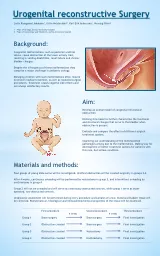
Slide37MeningoceleMyelomeningocele: Extension of CNS tissue through a defect in the vertebral column that occurs most commonly in the LS region.
-
Motor and sensory deficits in the lower extremities and problems with bowel and bladder control.
Slide38Slide39Neural Tube Defects Malformation of the anterior end of the neural tube- Anencephaly: Absence of the forebrain and the top of skull.
Slide40Neural Tube Defects Encephalocele: diverticulum of malformed CNS tissue extending through a defect in the cranium. - It most often involves the occipital region or the posterior
fossa
.
Slide41Forebrain MalformationsMegalencephaly : Abnormally large brain Microencephaly: Small brain, associated with a small head (
microcephaly
).
-
associated with chromosome abnormalities, fetal alcohol syndrome, and (HIV-1) infection acquired in
utero
.
Slide42Forebrain Malformations Disruption of neuronal migration and differentiation during development can lead to abnormalities of gyrationLissencephaly (agyria) : absent gyration leading to a smooth-surfaced brain.
Slide43- Polymicrogyria: increased number of irregularly formed gyri.
Slide44Holoprosencephaly : disruption of the normal midline patterning (incomplete separation of the two hemispheres)
Slide45Posterior Fossa AnomaliesThe Arnold-Chiari malformation (
Chiari
type II malformation) :
- Small posterior
fossa
- Misshapen midline cerebellum - Downward extension of the vermis through the foramen magnum - hydrocephalus and a lumbar myelomeningocele are also present.
Slide46Chiari type I malformation: - low-lying
cerebellar
tonsils that extend through the foramen magnum.
Dandy -Walker malformation
Slide47