
Hydatid disease in people is mainly caused by infection with the larval stage of the dog tapeworm Echinococcus granulosus It is an important pathogenic zoonotic and parasitic infection acquired from animals of humans following ingestion of tapeworm eggs excreted in the ID: 935633
Download Presentation The PPT/PDF document "Hydatid cyst disease Introduction" is the property of its rightful owner. Permission is granted to download and print the materials on this web site for personal, non-commercial use only, and to display it on your personal computer provided you do not modify the materials and that you retain all copyright notices contained in the materials. By downloading content from our website, you accept the terms of this agreement.


Slide1
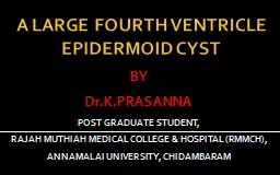
Hydatid cyst disease
Slide2Introduction
Hydatid
disease in people is mainly caused by infection with the larval stage of the dog tapeworm
Echinococcus
granulosus
. It is an important pathogenic,
zoonotic
and parasitic
infection (acquired from animals) of humans, following ingestion of tapeworm eggs excreted in the
faeces
of infected dogs.
Slide3Adult worm
Slide4Cystic
hydatid
disease usually affects the liver (50–70%) and less frequently the lung, the spleen, the kidney, the bones, and the brain
Echinococcus
granulosus
is spread almost all over t he world, especially in areas where sheep are raised, and is endemic in Asia, North Africa, South and Central America, North America, Canada and the Mediterranean region.
Slide5In many countries,
hydatid
disease is more prevalent in rural areas where there is a closer contact between people and dogs and various domestic animals which act as intermediate vectors.
Slide6Life cycle of hydatid disease
Slide7Layers of
hydatid
cyst
Pericyst
or adventitia
fibrous tissue induced by the expanding parasitic cyst
Th
e
ectocyst
or laminated layer
is elastic white covering, easily separable from the adventitia
.
Germinal layer
or
endocyst
is a single layer of cells lining the inner aspects of the
cyst and is the only living component, being responsible for the formation of the other layers .The
germinal layer produces clear fluid which attains a pressure of up to 300 mm of water, keeping the
endocyst
in intimate contact with the
pericyst
. The
endocyst
receives its
substance
from the
pericyst
.
Slide8Hepatic hydatid cyst (pathology and layers )
Slide9WHO CLASSIFICATION :
Group
1: Active group – cysts larger than 2 cm and often fertile.
Group 2: Transition group – cysts starting to degenerate
and entering
a transitional stage because of host resistance
or treatment
, but may contain viable
protoscolices
.
Group 3: Inactive group – degenerated, partially or totally
calcified cysts
; unlikely to contain viable
protoscolices
.
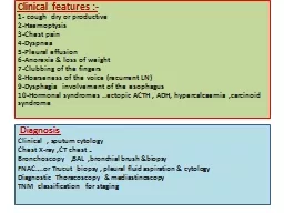
Slide10Clinical presentation :
The clinical features
are
highly variable. The spectrum of symptoms depends on the following:
Involved organs
Size of cysts and their sites within the affected organ or organs
Interaction between the expanding cysts and adjacent organ structures, particularly bile ducts and the vascular system of the liver
Symptoms due to pressure usually take a long time to manifest, except when they occur in the brain
.
Slide11Most symptomatic cysts are larger than 5 cm in diameter.
Bacterial infection of cysts and spread of
protoscolices
and larval material into bile ducts or blood vessels
Immunologic reactions such as asthma, anaphylaxis, or membranous nephropathy secondary to release of antigenic material
Slide12:clinical presentations (hepatic)
Hepatic hydatid cyst grow slowly and asymptomatic thus tend to be quite large at presentation .
Often discovered incidentally during diagnostic
w
ork up for un related complaint .
Symptomatic patients commonly complain of mild right upper quadrant (RUQ) pain and dyspepsia.
Slide13In case of sever abdominal pain it may indicate rupture ,
biliary
complication ,or secondary bacterial infection.
In case of fever with chills and jaundice indicate intra
biliary
rupture causing obstruction and
cholangitis
.
Uticaria
.
Slide14Complications of hepatic hydatid cyst
Rupture ……….. A-internal
B- external ….
intrabiliary
….
intrathoracic
….
intraperitoneal
Pressure effect;
e.g
obstructive jaundice
Secondary infection
Allergic reaction
A.urticatia
b-
brochospasm
c-Anaphylaxis
d –
eosinophilia
organ dysfunction :
cholangitis
,
biliary
cirrhosis .hepatic failure .
Spread ,recurrence .
Slide15Investigations
Laboratory
hematological test
:elevated total
leucocyte
count ,
esinophilia
.
Liver function test
.
Renal function test
.
Serological tests
:
(IHT),CFT, ELISA,IFAT.
Slide16Imaging studies
Plain x-ray
of abdomen :elevated right
hemdiaphragm
,calciftion of cyst.
C.X.R
: to exclude lung hydatid cyst.
Ultrasonograghic
study
The most important thing is the
CT scan
of abdomen .
ERCP,MRCP,MRI
:for
obstructive
jaundice,cholangitis
.
Slide17Ultrasound of the gall bladder (GB) and common bile
duct (CBD) showing a dilated CBD.
the
patient presented with jaundice from sludge in the CBD due
to daughter
cysts travelling down
biliary
channels in communication
with
cyst
.
Slide18Slide19Computerised
tomographic
(CT) scan of the upper
abdomen showing a
hypodense
lesion of the left lobe of the liver
Slide20CT scan showing liver
hydatid
cyst
(CT) scan showing a
hydatid
cyst of the pancreas.
Slide21Computerised
tomographic
(CT) scan showing disseminated hydatid cysts of the abdomen (a) and pelvis (b). The patient was started
on
albendazole
and lost to follow-up
Slide22Magnetic resonance cholangiopancreatography
(MRCP)
showing a large hepatic hydatid cyst with daughter cysts communicating
with the common bile duct causing obstruction and dilatation of the
entire
biliary
tree
Slide23Treatment of hydatid disease
Medical treatment (
antihelemthics
)
Indication ….. Concurrent pulmonary or disseminated disease.
….. Multiple
….. Recurrent
…… inaccessible
…..
Intaoperative
dissemination
…… unfit for surgery .
Slide24Albendazole
10-15mg/kg in 2divided doses for 28days .
Praziquantil
40mg /kg +
albedazole
Mebandazole
40-50 mg/kg for 6 to
12
months.
Slide25PAIR (puncture-aspiration-injection-reaspiration
)
Indication
for PAIR:
Refusal of surgery
Inoperable cases
Multiple cysts ≥5cm diameter in different liver segments .
Relapse post surgery .
Lack of response to chemotherapy
Slide26Slide27Contraindication to PAIR :
In accessible or hazardous location of cyst .
Dead in active cysts
Complications:
Urticatia
,fever ,
anaphylaxsis
,
biliary
fistula
subcpasular
haematoma
,hypotension shock.
Slide28Surgical treatment of hepatic hydatid disease
Indication for hepatic surgery :
Large cysts with suspected multiple cysts.
Superficial cysts with risk of spontaneous or post traumatic rupture .
Secondary bacterial infection of cyst .
Cystobiliary
communication
Pressure effects an adjacent .
Slide29Contraindication to hepatic surgery
In operable cases
Difficult access to cyst
Dead cyst
Surgical procedures for the treatment of hepatic
hydatid
disease:
open …….total or partial
pericystectomy
…….
Marsupialisation
and tube
drainge
with
omentoplasty
.
…….. Partial resection partial
hepatectomy
Laprascopic
precedure
Slide30Slide31Complication of hepatic
hydatid
surgery
Biliary
leak .
biliary
fistula .
Infection of residual cavity.
Cholangitis
Example of
scolicidal
agent :
20% hypertonic saline .
Absolute alcohol
Povidone
iodine .
Slide32Pulmonary hydatid disease
The lung is the second most common organ affected after the liver. The size of the cyst can vary from being very small to considerable size.
The right lung and lower lobes are slightly more often involved. The cyst is usually single.
The condition may be silent and found incidentally. Symptomatic patients present with cough, expectoration, fever, chest pain and sometimes
haemoptysis
. Silent cysts may present as an emergency due to rupture or an allergic reaction.
Slide33Uncomplicated cysts present as rounded or oval lesions on chest x-ray.
Erosion of the bronchioles results in air being introduced between the
pericyst
and the laminated membrane gives a fine radiolucent crescent
Slide34The mainstay of treatment of
hydatidosis
of the lung is surgery.
Medical treatment is less successful and considered when surgery is not possible because of poor general condition or diffuse
Slide35Thank you