
Spinal nerve and Reflex arc Dr Qazi Imtiaz Rasool OBJECTIVES Recall various components of somatic nervous system Explain structure of typical spinal nerve ID: 932342
Download Presentation The PPT/PDF document "Organization of Somatic Nervous system" is the property of its rightful owner. Permission is granted to download and print the materials on this web site for personal, non-commercial use only, and to display it on your personal computer provided you do not modify the materials and that you retain all copyright notices contained in the materials. By downloading content from our website, you accept the terms of this agreement.


Slide1
Organization of
Somatic Nervous system Spinal nerve and Reflex arc Dr. Qazi Imtiaz Rasool
Slide2OBJECTIVES
Recall various components of somatic nervous system. Explain structure of typical spinal nerve.Describe reflex arc.Identify clinical application.
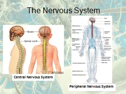
Slide3Nervous System
1.CNS 2.PNS 1. SOMATIC 2. AUTONOMIC 1. Brain 2.
Spinal
Cord
Slide4Somatic nervous system (SNS)
All parts of the nervous system outside of the brain and spinal cordSomatic System: Links spinal cord with body and sense organs; controls voluntary behaviorAutonomic System: Serves internal organs and glands; controls automatic functions such as heart rate and blood pressureEnteric System
Slide5Functional Classification
BRAINSPINAL CORD(CNS)PNSAFFERENTNERVESEFFERENTNERVESEXTERO-RECEPTORSINTERO-RECEPTORS
SOMATIC
AUTONOMIC
EFFECTOR
ORGANS
SKELETAL
MUSCLES
SMOOTH AND
CARDIAC MUSCLES
AND GLANDS
Slide6
Slide7Nerves Spinal nerves
Form lateral to intervertebral foramenWhere dorsal and ventral roots uniteThen branch and form pathways to destinationMotor nerves first branchWhite ramus Carries visceral motor fibers to sympathetic ganglion of autonomic nervous systemGray ramus Unmyelinated nerves , Return from sympathetic ganglion to rejoin spinal nervePeripheral Distribution of Spinal nerve
Slide8DRG
Afferent fiberEfferent fiber
Slide9Slide10Spinal Nerves
.Based on vertebrae where spinal nerves originate Positions of spinal segment and vertebrae change with ageCervical nerves Are named for inferior vertebra All other nerves Are named for superior vertebra
Slide11Peripheral Nerves
Epineurium wraps entire nerve Perineurium wraps fascicles of tractsEndoneurium wraps individual axons
Slide12Nerve structure
Nerves are only in the peripheryCable-like organs in PNS = cranial and spinal nervesConsists of 100-100,000 of myelinated + unmyelinated axons (nerve fibers)+ connective tissue + blood vesselsSupport Cells of the PNS Satellite cells ---Protect neuron cell bodies Schwann cells---Form myelin sheath
Slide13Morphology
of neuronTwo partsCell body (soma)ProcessesDendrites
Axon
1.membrane
2.perikaryon
3.nucleus
Presynaptic
terminals.
terminal (
bouton
/ button)
Slide14AXON
1.Plasmalemma--axolemma2.Cytoplasm--axoplasm
3, Axon
hillock;
Origin
4. No rough ER--No protein synthesis
5. Axon terminal
(
mitochondria,microtubues
,
Neurofilaments
,)
6.
Chromatophilic
-----
no
Nissl
body
Slide15FU
NCTIONAL PARTS OF AXON 1. Processes Integration zone2.Axon hillock 1ST portion of the axon plus the region of the cell body fro m which the axon leaves Neuron’s trigger zone
3.
Nerve
fiber
Single, elongated tubular extension that conducts AP away from the cell
Conducting zone of the neuron
4..Collaterals
Side branches of axon
5.Axon
terminals
Release
chemical
messengers other
cells
with
which they come into close
Output
zone of the neuron
Slide16REFLEX = reflection
is an involuntary, immediate, automatic andstereotyped response to a specific sensory stimulation.
Slide17Classification
CLINICALPHYSIOLOGICALNUMBER OF SYNAPSESSITEANATOMICALDEVELOPMENTFUNCTIONALON PURPOSESRESPONSE IS CONFINEDDEPENDING ON THE PART INVOLVEDCHARACTER OF THE RESPONSEOTHER REFLEXES
Slide18SIGNIFICANCE
HOMEOSTASIS (autonomic reflexes) TONE DURING RESTING STATETONE DURING TENSE MOTOR ACTIVITY3. POSTURE4. EQUILIBRIM5. EXECUTION OF MOVEMENTS6. SMOOTHNESS7. DAMPNESS during resting , walking, running, states8. ROLE AS PROPRIOCEPTOR( unconcouscious+ concious kinaesthetic sensations)
Slide19R-SIM
Reflex arc pathway R receptor neuron receives the stimuli2. S sensory neuron passes the impulse on3. I interneuron at the spinal cord processes4. M motor neuron acts
Slide20Simplified reflex arc
stimulus
Slide21stimulus
receptorSimplified reflex arc
Slide22stimulus
receptorsensory neurone
Simplified reflex arc
Slide23stimulus
receptorsensory neurone
spinal cord of central nervous system
Simplified reflex arc
Slide24stimulus
receptorsensory neurone
spinal cord of central nervous system
relay neurone
Simplified reflex arc
Slide25stimulus
receptorsensory neurone
spinal cord of central nervous system
relay neurone
motor neurone
Simplified reflex arc
Slide26stimulus
receptorsensory neurone
spinal cord of central nervous system
relay neurone
motor neurone
effector
Simplified reflex arc
Slide27stimulus
receptorsensory neurone
spinal cord of central nervous system
relay neurone
motor neurone
effector
response
Simplified reflex arc
Slide28Spinal Reflexes
Somatic reflexes mediated by the spinal cord are called spinal reflexesThese reflexes may occur without the involvement of higher brain centersAdditionally, the brain can facilitate or inhibit them
Slide29R 3 Inputs to Alpha
Motor Neurons29
(3) Spinal interneuron
DRG
(1) Afferent (sensory) neuron
(2) Upper
motor
neurons
Slide30Monosynaptic Reflexes
Slide31Stimulus
Biceps(flexor)contractsHandwithdrawnTriceps(extensor)relaxesAscending pathwayto brainResponseIntegrating center(spinal cord)Thermalpain receptor
in finger
Efferent pathway
Effector
organs
= Inhibitory interneuron= Excitatory interneuron= Synapse= Inhibits
= Stimulates
Afferent
Pathway
Slide32Efferent
pathway Afferentpathway Efferent pathway FlexormusclecontractsExtensormusclerelaxesFlexormusclerelaxesExtensormusclecontractsStimulusResponsePain
receptor
in heel
Injured
extremity(effectororgan)Integrating center
(spinal cord)Opposite
extremity(effectororgan)
Response
Slide33UMN lesions Weakness, paralysisSpasticity tendon reflexes +ve Babinski sign Little,if muscle atrophyNo fasiculation LMN lesionsweakness, paralysisflaccidity, hypotonia tendon reflexes -ve Babinski signMuscle
atrophy
F
asiculation
of muscle
Slide34UMN v LMN
Cortex
UMN
LMN
Muscle
Spasticity
Flaccidity
Slide35Reflex testing
0 = ABSENT1+ = HYPOREFLEXIA2+ = NORMAL3+ = HYPERREFLEXIA4+ = HYPERREFLEXIA & CLONUS
Slide36SPINAL SHOCK
Spinal shock is a state of transient physiological (rather than anatomical) reflex depression of cord function below the level of injury with associated loss of all sensorimotor functions. An initial increase in blood pressure is noted due to the release of catecholamines, followed by hypotension.
Slide37Shingles ( of the herpes family) In dorsal root ganglia and cranial nerves Initial infection: chicken pox virusPeripheral NeuropathyRegional loss of sensory or motor function Due to trauma or compression R metabolic causes