
BIOMEDICAL IMPORTANCE Besides water the diet must provide metabolic fuels mainly carbohydrates and lipids protein for growth and turnover of tissue proteins fiber for roughage minerals elements with specific metabolic functions and vitamins and essential fatty acids organic compounds ID: 931665
Download Presentation The PPT/PDF document "Digestion & Absorption" is the property of its rightful owner. Permission is granted to download and print the materials on this web site for personal, non-commercial use only, and to display it on your personal computer provided you do not modify the materials and that you retain all copyright notices contained in the materials. By downloading content from our website, you accept the terms of this agreement.


Slide1
Digestion & Absorption
Slide2BIOMEDICAL IMPORTANCE
Besides water, the diet must provide metabolic fuels (mainly carbohydrates and lipids), protein (for growth and turnover of tissue proteins), fiber (for roughage), minerals (elements with specific metabolic functions), and vitamins and essential fatty acids (organic compounds needed in small amounts for essential metabolic and physiologic functions).
The polysaccharides,
triacylglycerols
, and proteins that make up the bulk of the diet must be hydrolyzed to their constituent
monosaccharides
, fatty acids, and amino acids, respectively, before absorption and utilization. Minerals and vitamins must be released from the complex matrix of food before they can be absorbed and utilized.
Slide3Digestion of Carbohydrates:
The principal sites of dietary carbohydrate digestion are the mouth and intestinal lumen.
In the mouth:
During mastication, salivary α-amylase acts briefly on dietary starch and glycogen in a random manner, hydrolyzing some α(1→4) bonds.
Slide4Degradation of dietary glycogen by salivary or pancreatic α-amylase
Slide5In the stomach:
Carbohydrate digestion halts temporarily in the stomach, because the high acidity inactivates the salivary α-amylase.
in the small intestine:
When the acidic stomach contents reach the small intestine, they are neutralized by bicarbonate secreted by the pancreas, and pancreatic α-amylase continues the process of starch digestion. The final digestive processes occur at the mucosal lining of the upper jejunum, declining as they proceed down the small intestine, and include the action of several
disaccharidases
and
oligosaccharidases
.
Slide6isomaltase
cleaves the α(1→6) bond in
isomaltose
.
maltase cleaves maltose, both producing glucose.
sucrase
cleaves sucrose producing glucose and fructose.
lactase (β-
galactosidase
) cleaves lactose producing
galactose
and glucose. These enzymes are secreted through, and remain associated with, the luminal side of the brush border membranes of the intestinal mucosal cells.
Slide7Slide8Absorption of monosaccharides
The duodenum and upper jejunum absorb the bulk of the dietary sugars. Insulin is not required for the uptake of glucose by intestinal cells. However, different sugars have different mechanisms of absorption.
galactose
and glucose are transported into the mucosal cells by an active, energy-requiring process that requires a concurrent uptake of sodium ions; the transport protein is the sodium-dependent glucose
cotransporter
1 (SGLT-1).
Fructose uptake requires a sodium-independent monosaccharide transporter (GLUT-5) for its absorption.
Slide9Abnormal degradation of disaccharides
Slide10Digestive enzyme deficiencies:
Causes:
Hereditary deficiencies of
disaccharidases
.
Malnutrition.
drugs that injure the mucosa of the small intestine.
normal individuals with severe diarrhea lead to brush border enzymes are rapidly lost, causing a temporary, acquired enzyme deficiency. Thus, patients suffering or recovering from such a disorder cannot drink or eat significant amounts of dairy products or sucrose without exacerbating the diarrhea.
Slide11Lactose intolerance:
More
than three quarters of the world's adults are lactose intolerant. This is particularly manifested in certain races. For example, up to ninety percent of adults of African or Asian descent are lactase-deficient and, therefore, are less able to metabolize lactose than individuals of Northern European origin.
Slide12The mechanism
: by which this age-dependent loss of the enzyme occurs is not clear, but it is determined genetically and represents a reduction in the amount of enzyme protein rather than a modified inactive enzyme.
Treatment
:
for
this disorder is to reduce consumption of milk while eating yogurts and cheeses, as well as green vegetables such as broccoli, to ensure adequate calcium intake; to use lactase-treated products; or to take lactase in pill form prior to eating.
Slide13Abnormal lactose metabolism
Slide14Isomaltase-sucrase deficiency:
This enzyme deficiency results in an intolerance of ingested sucrose.
Treatment
: includes the withholding of dietary sucrose, and enzyme replacement therapy.
Slide15Diagnosis:
Identification of a specific enzyme deficiency can be obtained by performing oral tolerance tests with the individual disaccharides.
Measurement of hydrogen gas in the breath is a reliable test for determining the amount of ingested carbohydrate not absorbed by the body, but which is metabolized instead by the intestinal flora
.
Slide16DIGESTION & ABSORPTION OF LIPIDS:
The major lipids in the diet are
triacylglycerols
and, the remainder of the dietary lipids consists primarily of cholesterol,
cholesteryl
esters, phospholipids, and
unesterified
(“free”) fatty acids.
Slide17In the stomach:
The digestion of lipids begins in the stomach, catalyzed by an acid-stable lipase that originates from glands at the back of the tongue (lingual lipase). TAG molecules, particularly those containing fatty acids of short- or medium-chain length (less than 12 carbons, such as are found in milk fat), are the primary target of this enzyme. These same TAGs are also degraded by a separate gastric lipase, secreted by the gastric mucosa.
Slide18In
the small intestine:
emulsification of dietary lipids occurs in the duodenum. Emulsification increases the surface area of the hydrophobic lipid droplets so that the digestive enzymes, which work at the interface of the droplet and the surrounding aqueous solution, can act effectively.
Slide19TAG degradation
pancreatic enzymes:
pancreatic lipase :act on TAG molecules because they are too large to be taken up efficiently by the mucosal cells of the intestinal
villi
. which preferentially removes the fatty acids at carbons 1 and 3.
Colipase
:
also secreted by the pancreas, binds the lipase at a ratio of 1:1, and anchors it at the lipid-aqueous interface.
N.B:
Orlistat
, an
antiobesity
drug, inhibits gastric and pancreatic lipases, thereby decreasing fat absorption, resulting in loss of weight.
Slide20Cholesteryl ester degradation:
Most
dietary cholesterol is present in the free (
nonesterified
) form, with 10–15% present in the
esterified
form.
Cholesteryl
esters are hydrolyzed by pancreatic
cholesteryl
ester
hydrolase
(cholesterol esterase), which produces cholesterol plus free fatty acids.
Cholesteryl
ester
hydrolase
activity is greatly increased in the presence of bile salts.
Slide21Phospholipid degradation:
Pancreatic juice is rich in the
proenzyme
of
phospholipase
A
2
that, like
procolipase
, is activated by
trypsin
and, like
cholesteryl
ester
hydrolase
, requires bile salts for optimum activity.
Phospholipase
A
2
removes one fatty acid from carbon 2 of a
phospholipid
, leaving a
lysophospholipid
. The remaining fatty acid at carbon 1 can be removed by
lysophospholipase
, leaving a
glycerylphosphoryl
base.
Slide22Control of lipid digestion:
Cholecystokinin
(CCK, formerly called
pancreozymin
):
a small peptide hormone is produced by the Cells in the mucosa of the jejunum and lower duodenum in response to the presence of lipids and partially digested proteins entering these regions of the upper small intestine.
CCK
acts on the gallbladder (causing it to contract and release bile—a mixture of bile salts, phospholipids, and free cholesterol), and on the exocrine cells of the pancreas (causing them to release digestive enzymes
).
It also decreases gastric motility, resulting in a slower release of gastric contents into the small intestine
.
Slide23Secretin
,
in response to the low pH of the
chyme
entering the intestine.
Secretin
causes the pancreas and the liver to release a watery solution rich in bicarbonate that helps neutralize the pH of the intestinal contents, bringing them to the appropriate pH for digestive activity by pancreatic enzymes.
Slide24Absorption of lipids by intestinal mucosal cells (enterocytes
)
Free fatty acids, free cholesterol, and 2-monoacylglycerol are the primary products of lipid digestion in the jejunum. These, plus bile salts and fat-soluble vitamins, form mixed micelles—disk-shaped clusters of
amphipathic
lipids that coalesce with their hydrophobic groups on the inside and their hydrophilic groups on the outside. Mixed micelles are, therefore, soluble in the aqueous environment of the intestinal lumen.
Slide25Absorption of lipids contained in a mixed micelle by an intestinal mucosal cell.
Slide26Resynthesis
of TAG and
cholesteryl
esters
The
mixture of lipids absorbed by the
enterocytes
migrates to the endoplasmic reticulum where biosynthesis of complex lipids takes place.
Slide27Slide28Digestion of Dietary Proteins
Most of the nitrogen in the diet is consumed in the form of protein, Proteins are generally too large to be absorbed by the intestine. They must, therefore, be hydrolyzed to yield their constituent amino acids, which can be absorbed.
Proteolytic
enzymes responsible for degrading proteins are produced by three different organs: the stomach, the pancreas, and the small intestine .
Slide29Digestion of proteins by gastric secretion
The digestion of proteins begins in the stomach, which secretes gastric juice—a unique solution containing hydrochloric acid and the
proenzyme
,
pepsinogen
.
Slide30Hydrochloric acid:
Stomach acid is too dilute (pH 2–3) to hydrolyze proteins. The acid functions instead to kill some bacteria and to denature proteins, thus making them more susceptible to subsequent hydrolysis by proteases.
Pepsin:
This acid-stable
endopeptidase
is secreted by the serous cells of the stomach as an inactive
zymogen
(or
proenzyme
),
pepsinogen
.
Pepsinogen
is activated to pepsin, either by
HCl
, or
autocatalytically
by other pepsin molecules that have already been activated. Pepsin releases peptides and a few free amino acids from dietary proteins.
Slide31Digestion of proteins by pancreatic enzymes
Release of zymogens:
The release and activation of the pancreatic zymogens is mediated by the secretion of
cholecystokinin
and
secretin
, two polypeptide hormones of the digestive tract.
Enteropeptidase
(formerly called
enterokinase
)— an enzyme synthesized by and present on the luminal surface of intestinal mucosal cells of the brush border membrane—converts the pancreatic
zymogen
trypsinogen
to
trypsin
.
Trypsin
subsequently converts other
trypsinogen
molecules to
trypsin
by cleaving a limited number of specific peptide bonds in the
zymogen
.
ens
.
Slide32Celiac disease (celiac
sprue
)
is a disease of
malabsorption
resulting from immune-mediated damage to the small intestine in response to ingestion of gluten, a protein found in wheat and other grains.
Digestion of
oligopeptides
by enzymes of the small intestine
The luminal surface of the intestine contains
aminopeptidase
—an
exopeptidase
that repeatedly cleaves the N-terminal residue from
oligopeptides
to produce free amino acids and smaller peptides.
Slide33Absorption of amino acids and dipeptides
Free
amino acids are taken into the
enterocytes
up by a Na
+
-linked secondary transport system. Di- and
tripeptides
, however, are taken up by a H
+
-linked transport system. There, the peptides are hydrolyzed in the
cytosol
to amino acids before being released into the portal system. Thus, only free amino acids are found in the portal vein after a meal containing protein. These amino acids are either metabolized by the liver or released into the general circulation.
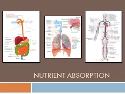
Slide34DIGESTION & ABSORPTION OF VITAMINS & MINERALS
Vitamins and minerals are released from food during digestion—though this is not complete—and the availability of vitamins and minerals depends on the type of food and, especially for minerals, the presence of chelating compounds.
Slide35Calcium Absorption Is Dependent on Vitamin D
vitamin
D is required for the intestinal absorption of calcium. Synthesis of the intracellular
calcium binding
protein,
calbindin
,
required for calcium absorption, is induced by vitamin D, which also affects the permeability of the mucosal cells to calcium, an effect that is rapid and independent of protein synthesis
.
Phytic
acid (
inositol
hexaphosphate
) in cereals binds calcium in the intestinal lumen, preventing its absorption.
Slide36Other minerals, including zinc, are also
chelated
by
phytate
. This is mainly a problem among people who consume large amounts of unleavened whole wheat products; yeast contains an enzyme,
phytase
,
which
dephosphorylates
phytate
, so rendering it inactive.
High concentrations of fatty acids in the intestinal lumen—as a result of impaired fat absorption—can also reduce calcium absorption by forming insoluble calcium salts; a high intake of oxalate can sometimes cause deficiency, since calcium oxalate is insoluble.
Slide37Iron Absorption
Absorption of iron is strictly regulated. Inorganic iron is
accumulatedin
intestinal mucosal cells bound to an intracellular protein,
ferritin
.
. Iron can only leave the mucosal cell if there is
transferrin
in plasma
.
only about 10% of dietary iron is normally absorbed and only 1–5% from many plant foods
.
Inorganic iron is absorbed only in the Fe
2+
(reduced) state, and for that reason the presence of reducing agents will enhance absorption.