

MD Hom Prof amp HoD Department of Pathology Definition Edema is an abnormal accumulation of fluid in the cavities and intercellular spaces of the body Oedema Primary factors favoring edema are increased capillary hydrostatic pressure increased venous pressure decreas ID: 929988
Download Presentation The PPT/PDF document "edema Dr. R. S. Gopika" is the property of its rightful owner. Permission is granted to download and print the materials on this web site for personal, non-commercial use only, and to display it on your personal computer provided you do not modify the materials and that you retain all copyright notices contained in the materials. By downloading content from our website, you accept the terms of this agreement.


Slide1
edema
Dr.
R. S.
Gopika
M.D. (
Hom
)
Prof.
&
HoD
Department of Pathology
Slide2Definition
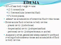
Edema is an abnormal accumulation of fluid in the cavities and intercellular spaces of the body.
Slide3Oedema
. Primary factors favoring edema are increased capillary hydrostatic pressure (increased venous pressure), decreased osmotic pressure of plasma (hypoproteinemia
), decreased tissue tension and lymphatic drainage, increased osmotic pressure of tissue fluids, and increased capillary permeability. Additional renal and hormonal factors are important. Clinical manifestations may consist of a steady weight gain or localized or generalized swelling.
Slide4Slide5Slide6Slide7Oedema ….cont
Two forces are responsible for maintaining the fluid in specific areas or ‘pulling’ and ‘pushing’ fluid into other areas. These forces are known as
hydrostatic pressure and osmotic pressure. Hydrostatic pressure is the force that pushes fluid from an area of high pressure to low pressure.
Osmotic pressure is the force that draws fluid from an area of low electrolyte concentration to one of a higher electrolyte concentration.
Slide8Slide9Other mechanism which influence the movement of fluid with in the body.
Slide10Other factors …cont
Slide11PATHOPHYSIOLOGY OF EDEMA FORMATION
There are two basic steps involved in edema formation:An alteration in capillary hemodynamics
that favors the movement of fluid from the vascular space into the interstitium.The retention of dietary or intravenously administered sodium and water by the kidneys
Slide12Causes of Edema
Edema will occur under these circumstances :Increased hydrostatic pressure will push fluid out of the vessels into tissue spaces. This results in edema.
Reduced osmotic pressure within the vessels will not pull fluid from the tissue spaces into the vessel. The fluid accumulates within the tissue space and results in edema.Fluid retention (
water retention
) where there is excessive fluid within the blood vessel and tissue spaces. If the body is not able to pass out this excess fluid, it will be retained within the tissue spaces thereby resulting in edema.
Slide13Increased vascular permeability
is when blood vessel wall allows fluid to pass out of the blood vessel unabated. Fluid from the tissue spaces are not drawn into the blood vessel fast enough and fluid remains in the tissue space thereby resulting in edema.Lymphatic obstruction is where the lymph vessels are blocked at some point and the interstitial fluid cannot be drained from the tissue spaces. Fluid accumulates in the tissue space and the result is edema.
Slide14Increased Hydrostatic Pressure
Impaired venous return Congestive heart failure Constrictive
pericarditis Ascites (liver cirrhosis) Venous obstruction or compression
Thrombosis
External pressure (e.g., mass)
Lower extremity inactivity with prolonged dependency Arteriolar dilation Heat
Neurohumoral
dysregulation
Slide15Slide16Reduced Plasma Osmotic Pressure/ Hypoproteinemia
Protein-losing glomerulopathies (
nephrotic syndrome) Liver cirrhosis (ascites) Malnutrition
Protein-losing
gastroenteropathy
Slide17Lymphatic Obstruction
Inflammatory Neoplastic Postsurgical Postirradiation
Slide18Sodium Retention
Excessive salt intake with renal insufficiency Increased tubular reabsorption of sodium Renal
hypoperfusion Increased renin-angiotensin-aldosterone secretion
Slide19Inflammation
Acute inflammation Chronic inflammation Angiogenesis
Slide20Pathogenesis
Sequence of events leading to systemic edema due to primary heart failure, primary renal failure, or reduced plasma osmotic pressure (as in malnutrition, diminished hepatic protein synthesis, or loss of protein owing to the
nephrotic syndrome).
Slide21Slide22Changes
Microscopical tissue cells or fibers are seperated
intercellular space widened fluid is homogenous or granular
Slide23Macroscopical
Tissue, organ swollen, boggy, gelatinousOn cutting exude fluidLoss of elasticity of tissues- pitting
Slide24Edema Terminology
There are different medical terms for edema in specific areas or organs of the body.Anasarca is the term for severe generalized edema.
Ascites is the term for excessive fluid accumulation within the peritoneal cavity. This is the area between the lining of the abdomen and organs within the abdominal cavity.Pleural effusion is the term for edema in the pleural space between the outer layers of the lung. It is also known as a hydrothorax.
Slide25Pericardial effusion
is the term for edema within the pericardial space between the outer layers of the heart. It is also known as a hydropericardium.Pulmonary edema
is the term for edema within the lungs.Cerebral edema is the term for edema within the brain.Lymphedema is the excessive fluid accumulation within tissues because the tissue fluid cannot be drained by the lymphatic vessels in that area.
Slide26Hepatic edema
is the term for excessive fluid accumulation in tissues due to a liver dysfunction.Cardiac edema is the term for excessive fluid accumulation in tissues due to heart failure.Renal edema
is the excessive fluid accumulation in they body’s tissues due to kidney disease or failure.
Slide27Morphology
Edema is most easily recognized grossly; microscopically, edema fluid is reflected primarily as a clearing and separation of the extracellular matrix elements with subtle cell swelling. Although any organ or tissue in the body may be involved, edema is most commonly encountered in subcutaneous tissues, lungs, and brain.
Slide28Subcutaneous edema
can be diffuse or more prominent in regions with high hydrostatic pressures; the ultimate distribution depends on the underlying etiology. Even diffuse edema is usually more prominent in certain body areas as a result of the effects of gravity; a gravity-dependent distribution is referred to as
dependent edema Dependent edema is a prominent feature of cardiac failure, particularly of the right ventricle
Slide29Finger pressure over significantly edematous subcutaneous tissue displaces the interstitial fluid and leaves a finger-shaped depression, so-called
pitting edema.
Slide30oedema
is most commonly caused by:Physical inactivity - edema is more prevalent among people who do not exercise at all, and walk very little.
Standing or sitting still for long - if you stand or sit still for a long time there is a much higher chance of swelling.
Slide31High altitudes
- especially when combined with physical exertion. Acute mountain sickness can lead to high altitude pulmonary edema or high altitude cerebral edema.Heat - especially when combined with physical exertion. During high temperatures the body is less efficient at removing fluid from tissues, especially around the ankles.
Burns - the skin reacts to a burn by retaining fluid, causing localized swelling
Slide32Pregnancy
- during pregnancy the woman releases hormones which encourage the body to retain fluids. Pregnant women tend to retain much more sodium and water than women who are not pregnant. When the woman is resting in a reclined position the enlarged uterus occasionally compresses the inferior vena cava, causing obstruction of both femoral veins, leading to edema
A pregnant woman's blood is hypercoaguble (clots more easily), raising the risk of deep venous thrombosis (DVT), a cause of edema
Slide33Menstruation and pre-menstruation
- hormone levels fluctuate during the menstrual cycle. During the days before menstrual bleeding there will be a reduction in the levels of the hormone progesterone, which may cause fluid retention.The contraceptive pill
- estrogen can cause fluid retention.Menopause - around the period of the menopause as well as after it, hormone fluctuations can cause fluid retention.
Slide34Malnutrition and/or bad diet
- low consumption of thiamine (vitamin B1), as well as insufficient vitamins B6 and B5 may contribute toward fluid retention. Low levels of albumin levels may also play a part - low albumin levels can also be caused by kidney disease.
Slide35Edema can also be caused by the following diseases:
Kidney disease/damage - patients with kidney disease may not be able to eliminate enough fluid and sodium from the blood. This results in more pressure on the blood vessels, which causes some of the liquid to leak out.
Kidney disease patients with edema will generally have swelling around their legs and eyes
Slide36Renal oedema
Damage to the capillaries in the kidneys (glomeruli) that filter waste and excess fluids from the blood can result in nephrotic
syndrome. Among the many symptoms of nephrotic syndrome is an insufficient level of blood albumin, which leads to edema.
Slide37Heart failure
this is when the heart cannot pump blood properly to all parts of the body. If one or both of the lower chambers of the heart lose the ability to pump blood effectively, the blood can accumulate in the limbs, causing edema
Slide38Chronic lung disease -
this includes many lung diseases, such as asthma, chronic bronchitis COPD, emphysema, pulmonary fibrosis and sarcoidosis
. Some patients may experience an accumulation of fluids in the lungs - pulmonary edema.
Slide39Liver disease
such as cirrhosis, which causes scarring of the liver. This affects liver function, which causes the secretion of hormones and fluid-regulating chemicals to change. People with cirrhosis of the liver also have increased pressure within the portal vein
The problems can lead to fluid retention in the legs and ascites (abdominal cavity).
Slide40Cardiac edema
Slide41Cardiac oedema
Generalised oedemaStarts in most dependent parts
Slide42When the heart ceases to meet the requirements of the body for oxygen, the sympathetic-adrenal system is activated.
This occurs in people with a healthy heart when the demands for oxygen are excessive—for example, in heavy muscular work—and in subjects with a failing heart when the demands are normal or small. Eventually, when the heart is unable to meet even the ordinary requirements of everyday life, the sympathetic activity becomes more or less continuous.
It may lessen during rest at night, but with a further failing of the heart its output may become inadequate even in complete rest.
Slide43Angioneurotic
oedema Angioedema or
Quincke's edema is the rapid swelling (edema) of the dermis, subcutaneous tissue, mucosa and submucosal tissues. It is very similar to urticaria
, but
urticaria
, commonly known as hives, occurs in the upper dermis.
The term
angioneurotic
oedema
was used for this condition in the belief that there was nervous system involvement but this is now thought not to be the case
Slide44Slide45Types
Acquired angioedema is usually caused by allergy and occurs together with other allergic symptoms and urticaria. It can also happen as a side-effect to certain medications, particularly ACE inhibitors.
Hereditary angioedema (HAE) - caused by a genetic mutation that is inherited in an autosomal dominant form.
Slide46Pathophysiology
Bradykinin plays a critical role in all forms of hereditary
angioedema. This peptide is a potent vasodilator and increases vascular permeability, leading to rapid accumulation of fluid in the interstitium. This is most obvious in the face, where the skin has relatively little supporting connective tissue, and edema develops easily.
Various mechanisms that interfere with
bradykinin
production or degradation can lead to
angioedema
Slide47In
hereditary angioedema, bradykinin formation is caused by continuous activation of the complement system due to a deficiency in one of its prime inhibitors, C1-esterase (aka: C1-inhibitor or C1INH), and continuous production of
kallikrein. Consumption of foods which are themselves vasodilators such as alcohol or cinnamon can increase the probability of an angioedema episode in susceptible patients
Slide48Pulmonary edema
is fluid accumulation in the lungs.It leads to impaired gas exchange and may cause respiratory failure. It is due to either failure of the heart to remove fluid from the lung circulation ("
cardiogenic pulmonary edema") or a direct injury to the lung parenchyma ("noncardiogenic pulmonary edema").
Slide49Pulmonary edema with small pleural effusions on both sides.
which shows increased fluid in the
alveolar walls.
Kerley
B lines
, increased
vascular filling ,
pleural effusions
,
upper lobe diversion (increased
blood flow to the higher parts of the lung)
Slide50Cardiogenic
Congestive heart failureSevere heart attack with left ventricular failure
Severe arrhythmias (tachycardia/fast heartbeat or bradycardia/slow heartbeat)Hypertensive crisisPericardial effusion with tamponade
Fluid overload, e.g., from kidney failure or intravenous therapy
Slide51Non-cardiogenic
May occur after upper airway obstruction, intravenous fluid overload, neurogenic
causes (seizures, head trauma, strangulation, electrocution). Can also be seen with ARDS (acute respiratory distress syndrome).
Slide52Alveolar
Inhalation of toxic gasesPulmonary contusion, Aspiration
Reperfusion injuryMultiple blood transfusionsSevere infection
Slide53Acute pulmonary oedema
left vent out put pul
venous pr fluid in alveolar walls fluid in alveolar spaces lung compliance
impaired gas exchange pink frothy sputum
Slide54Cerebral oedema
Slide55Slide56Thank you