
Uncomplicated Crown Fracture Primary tooth Smooth sharp edges Followup 1 wk 4 wk 68 wk 3 mo 4 mo 6 mo 1 yr Yearly Enamel fracture No F U recommended Enameldentin fracture ID: 929864
Download Presentation The PPT/PDF document "Enamel fracture Enamel-Dentine facture" is the property of its rightful owner. Permission is granted to download and print the materials on this web site for personal, non-commercial use only, and to display it on your personal computer provided you do not modify the materials and that you retain all copyright notices contained in the materials. By downloading content from our website, you accept the terms of this agreement.


Slide1
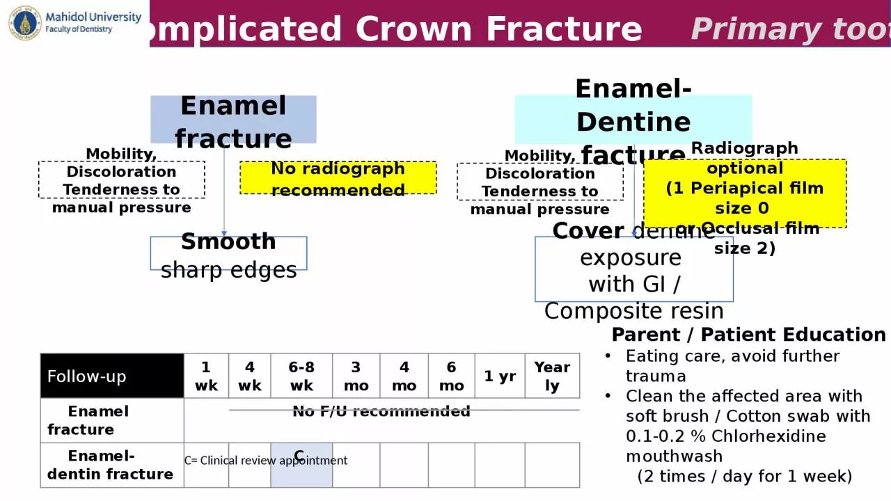
Enamel fracture
Enamel-Dentine facture
Uncomplicated Crown Fracture
Primary tooth
Smooth
sharp edges
Follow-up1 wk4 wk6-8 wk3 mo4 mo6 mo1 yrYearly Enamel fractureNo F/U recommended Enamel-dentin fractureC
Cover dentine exposure with GI / Composite resin
No radiograph recommended
Radiograph optional(1 Periapical film size 0 or Occlusal film size 2)
Mobility, DiscolorationTenderness to manual pressure
Mobility, DiscolorationTenderness to manual pressure
Parent / Patient Education
Eating care, avoid further trauma
Clean the affected area with soft brush
/ Cotton swab with 0.1-0.2 % Chlorhexidine mouthwash (2 times / day for 1 week)
C= Clinical review appointment
Slide2Complicated crown fracture
Complicated Crown Fracture
Primary tooth
1 Periapical film size 0 or Occlusal film size 2
+
Soft tissue radiograph
Mobility, DiscolorationTenderness to manual pressureLarge pulp exposurePartial pulpotomy withCa(OH)2 or Biodentine + RestorationSmall pulp exposureCervical pulpotomy withCa(OH)2 or Biodentine + RestorationFollow-up1 wk4 wk6-8 wk3 mo4 mo6 mo1 yrYearly Complicated crown fracture
C
C
C+R
Local anesthesia
Local anesthesia
Parent/ Patient Education
Eating care, avoid further trauma
Clean the affected area with soft brush
/
Cotton swab with 0.1-0.2 % Chlorhexidine mouthwash
(2 times
/
day for 1 week)
C = Clinical review appointment R = Radiographic advised
Slide3Crown-root fracture
Fracture
Primary tooth
Remove loose fragment
Restorable
Unrestorable
No pulp exposedCover dentine with GIPulp exposedPulpotomy / Pulpectomy(Depend on stage of root development)Crown-Root Fracture1 Periapical film size 0 or Occlusal film size 2Mobility, DiscolorationTenderness to manual pressureLocal anesthesiaExtract loose fragment and leave any firm rootFollow-up1 wk4 wk6-8 wk3 mo4 mo6 mo
1 yrYearly
Crown-Root fracture
C
C
C+R
Parent / Patient Education
Eating care, avoid further trauma
Clean the affected area with soft brush
/
Cotton swab with 0.1-0.2 % Chlorhexidine mouthwash
(2 times
/
day for 1 week)
C = Clinical review appointment R = Radiographic advised
Slide4Root fracture
Fracture
Primary tooth
Coronal segment is displaced.
Follow-up
1
wk4 wk8 wk1 yrYearlyCC + SC C C*Excessively mobile & Occlusal interferenceNot excessively mobileExtract coronal fragmentLeave apical fragmentReposition coronal fragmentIf unstable Splint 4 weeksCoronal segment is not displaced.No treatmentLeave spontaneous repositionEven if there is some occlusal interference. Follow-up
1 yr
Yearly
C
C*C* = Clinical follow-up yearly until eruption of permanent teeth
Follow-up
1
wk
6-8
wk1
yr
Yearly
C
C
C
C*
Root
Fracture
Mobility, Discoloration
Tenderness to manual pressure
Local anesthesia
1 Periapical film size 0 or Occlusal film size 2
Parent / Patient Education
Eating care, avoid further trauma
Clean the affected area with soft brush
/
Cotton swab with 0.1-0.2 % Chlorhexidine mouthwash
(2 times
/
day for 1 week)
C = Clinical review appointment
S
= Splint removal
Slide5Alveolar fracture
Reposition and Splint 4 weeks
C** = Further follow-up at 6 years old: monitor eruption of permanent teeth)
Follow-up
1
wk
4 wk8 wk3 mo4 mo6 mo1 yrYearly6 yrAlveolar fractureCC+R+SC C +RC**Alveolar FracturePrimary toothMobility, DiscolorationTenderness to manual pressure
Local anesthesia
1 Periapical film size 0 or Occlusal film size 2
Monitor eruption of permanent teeth at 6 years of age (C**)A lateral radiograph may give information about the relationship between the maxillary and mandibular dentitions
and if the segment is displaced in a labial directionIf Parent / Patient Education
Eating care, avoid further traumaClean the affected area with soft brush / Cotton swab with 0.1-0.2 % Chlorhexidine mouthwash (2 times / day for 1 week)
C = Clinical review appointment R = Radiographic advised S
= Splint removal
Slide6Concussion
Subluxation
Concussion & Subluxation
Primary tooth
Observe
Follow-up
1 wk4 wk6-8 wk3 mo4 mo6 mo1 yrYearly ConcussionCCSubluxationCC
C*
Observe
C*= Clinical follow-up yearly until eruption of permanent teeth
Mobility, Discoloration
Tenderness to manual pressure
Mobility, Discoloration
Tenderness to manual pressure
No radiograph recommended
1 Periapical film size 0 or Occlusal film size 2
Parent / Patient Education
Eating care, avoid further trauma
Clean the affected area with soft brush
/
Cotton swab with 0.1-0.2 % Chlorhexidine mouthwash
(2 times
/
day for 1 week)
C = Clinical review appointment
Slide7Extrusive luxation
Follow-up
1
wk
4
wk
6-8 wk3 mo4 mo6 mo1 yrYearlyExtrusionCC C C*Extrusive LuxationNot interfere occlusionExcessively mobile / Extrude > 3 mm.Leave spontaneous reposition Extraction Primary tooth
1 Periapical film size 0 or Occlusal film size 2
Mobility, DiscolorationTenderness to manual pressure
Local anesthesia
C*= Clinical follow-up yearly until eruption of permanent teeth
Parent / Patient Education Eating care, avoid further traumaClean the affected area with soft brush / Cotton swab with 0.1-0.2 % Chlorhexidine mouthwash (2 times
/ day for 1 week)
C = Clinical review appointment
Slide8Lateral luxation
Follow-up
1
wk
4
wk
8 wk3 mo4 mo6 mo1 yrYearlyLateral luxationCC+SC CC C*Lateral luxationMinimal / No occlusal interferenceSevere displacementAllows spontaneous reposition (usually 6 months)Extraction
Reposition &Splint for 4 weeks
Primary tooth
1 Periapical film size 0 or Occlusal film size 2Mobility, DiscolorationTenderness to manual pressure
C*= Clinical follow-up yearly until eruption of permanent teeth
Local anesthesia
Parent/ Patient Education
Eating care, avoid further trauma
Clean the affected area with soft brush
/ Cotton swab with 0.1-0.2 % Chlorhexidine mouthwash (2 times /
day for 1 week)
C = Clinical review appointment
S
= Splint removal
Slide9Intrusive luxation
Follow-up
1
wk
4
wk
6-8 wk3 mo4 mo6 mo1 yrYearly6 yrIntrusionCC CCC**AvulsionC
C**
Intrusive Luxation & Avulsion
Primary tooth
Allow spontaneous reposition
(usually 6-12 months)
Avulsion
No replantation
C= Clinical review appointment
C**= Further follow-up at 6 years old: monitor eruption of permanent teeth
For severe intrusion
,
monitor eruption of permanent teeth at 6 years old (C**)
1 Periapical film size 0 or Occlusal film size 2
Mobility, Discoloration
Tenderness to manual pressure
1 Periapical film size 0
or Occlusal film size 2
Monitor eruption of permanent teeth at 6 years old (C**)
Parent / Patient Education
Eating care, avoid further trauma
Clean the affected area with soft brush
/
Cotton swab with 0.1-0.2 % Chlorhexidine mouthwash
(2 times
/
day for 1 week)