
Grams Negative Bacilli Non enteric bacilli Enteric bacilli Latelactose fermenting LLF NLF after 24h LF after 48h E coli Escherichia coli Klebsiella species Enterobacter species ID: 929462
Download Presentation The PPT/PDF document "Lactose fermenting (LF) Non-lactose ferm..." is the property of its rightful owner. Permission is granted to download and print the materials on this web site for personal, non-commercial use only, and to display it on your personal computer provided you do not modify the materials and that you retain all copyright notices contained in the materials. By downloading content from our website, you accept the terms of this agreement.


Slide1
Lactose fermenting (LF)
Non-lactose fermenting (NLF)
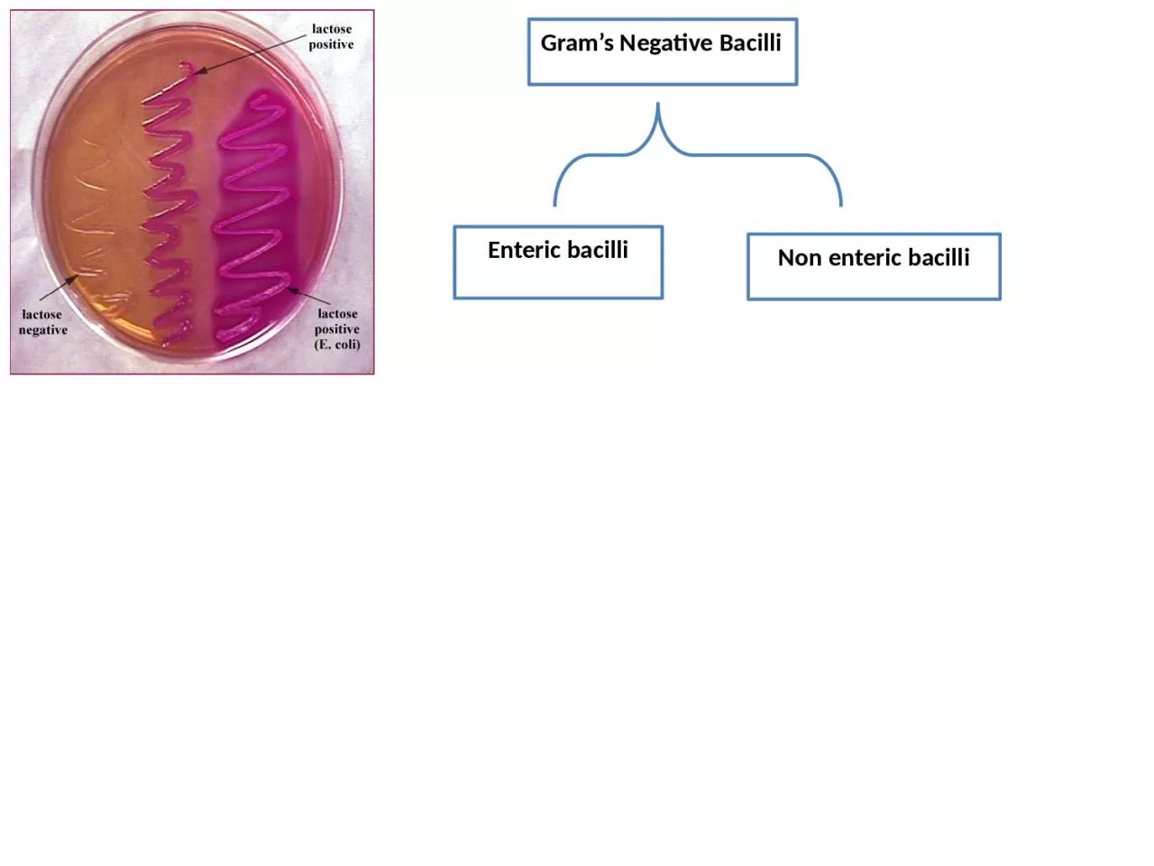
Gram’s Negative Bacilli
Non enteric bacilli
Enteric bacilli
Late-lactose fermenting (LLF): NLF after 24h, LF after 48h.
E. coli (Escherichia coli)
.
Klebsiella species
.
Enterobacter species.
Citrobacter
Serratia
Others
e.g. Salmonella, Shigella,
proteus,Yersinia
.
Slide2Non lactose fermenting
lactose fermenting
Slide3Family:
Enterobacteriaceae
(ENTERIC BACILLI, COLIFORM).
GENERAL CHARACTERISTICS
* G – ve bacilli or coccobacilli. * Aerobic or Facultative anaerobes.
* Ferment glucose and produce acid + gas.
* Oxidase –ve, catalase +ve * Most of them are normal flora of
human & animal GIT. * Grow well on MacConkey’s agar.
* Transmission either endogenous or person to person
especially in hospital.
Slide4E. coli (Escherichia coli)
Normal flora of human and animal GIT, human female genital tract.
Under electron microscope
Slide5E. coli (Escherichia coli)
There are many
STRAINS SEROTYPED
according to
O
Ag, H Ag, K Ag.
Most strains are Motile, un-capsulated.
Produced dry colonies.
Slide6E. coli (Escherichia coli) infections
DISEASES
INTESTINAL infections
EXTRAINTESTINAL infections
Slide7E. coli (Escherichia coli) infections
INTESTINAL infections
:(caused by some strains)
(e.g. Traveler's diarrhea, dysentery, hemorrhagic infection
).Source of infection either human or animal product.
E. coli
serotypes
Enterotoxigenic
E.coli
(ETEC)
Enteropathogenic
E.coli (EPEC)
Enteroinvasive E.coli (EIEC) Enterohemorrhagic E.coli (EHEC)
Enteroadhesive E. coli (EAEC)
Slide82) EXTRAINTESTINAL infections
Wound infections, Pneumonia, Bacteremia, Septicemia, UTI .
Its the
common cause of G-ve nosocomial
infection.E. coli (Escherichia coli) infections
Slide9Klebsiella
spp
Klebsiella pneumoniae,
K.oxytoca
, K.ozaenae
Normal flora of GIT.
Non - motile.
Capsulated by large capsule. Mucoid colonies.
Causes
Necrotizing Pneumonia,
Nosocomail infections like UTI
mainly in debilitated patients.
Slide10Laboratory Diagnosis
1
) Specimens
(site of infection e.g. urine, blood, pus…etc).
2) Staining Gram’s stain: Enterobacteriaceae resemble each other morphologically under microscope. So it is difficult to diagnose by microscope.
Slide11Staining
Capsular stain:
Klebsiella stained by this stain, bacilli surrounded by hallo zone.
Laboratory Diagnosis
Slide12Laboratory Diagnosis
3) Culture:
37C°, 24-48h
A) Differential media:
MacConkey’s agar
(selective and differential media):
A.) Lactose fermenter (Pink colonies after 24 hrs.):
E. coli, Klebsiella,
Enterobacter.B.) Late lactose fermenter (Pink after 48 hrs.): Serratia, Citrobacter
. C.) Non – lactose fermenter (Pale colonies after 24 or 48 hrs.): Proteus
, Salmonella
, Shigella.
Slide13MacConkey agar
(selective and differential media)
Bile salt:
Inhibit G-ve other than Enterobacteriaceae
Crystal violet: Inhibit G
+
ve bacteriaNeutral
red: Fermentation indicator Lactose:
Differential between genera Nutrient agar
Laboratory Diagnosis
Slide14Laboratory Diagnosis
E.coli
on MacConkey’s agar
(pink colony due to lactose fermentation
Dry, discreet pink colonies
Slide15Laboratory Diagnosis
Klebsiella
on MacConkey’s agar
(pink mucoid colony due to lactose fermentation
Slide162) EMB (Eosin Methylene Blue)
contain special dye:
E.coli
colonies appears green metallic sheen
Others
no green metallic sheen colonies or colorless.
Laboratory Diagnosis
E.coli
on EMB
Slide17Laboratory Diagnosis
Slide18Laboratory Diagnosis
B) Non differential medium:
Like Blood agar
Enterobacteriaceae appears (
large, gray, smooth)
Slide194) Biochemical tests :
1- (
IMViC
) test: used to differentiate between E. coli
and Klebsiella.
I
ndole:- Tryptophan (a.a) Tryptophanase Indole
add Kovac’s reagent
red ring (+ve).
M
ethyl red:- Glucose phosphate broth (fer
.) Acid (
decrease PH)
Methyl red red
(+ ve), yellow (-ve) Voges
proskauer:- Glucose ph. broth (fermentation) acetyl methyl carbinol
(5% α – naphthol) + (40% KOH) dark brown (+ve), (-ve) brown-greenC
itrate:
Simmon
Citrate agar(utilize citrate) citrase
Blue (+ ve),
Green
(- ve). Media contains bromthymol blue indicator.
Slide204) Biochemical tests :
1- (
IMViC
) test: used to differentiate between
E. coli and Klebsiella
.
Laboratory Diagnosis
Bacteria
I
M
V
C
E. coli
+ (red ring)+ (
red) - (yellow)- (
green) With growthKlebsiella- (colorless ring)
- (yellow)+ (red)
+ (blue)
Without growth
Slide21Laboratory Diagnosis
Indole
test
Slide22Laboratory Diagnosis
Methyl red test
Slide23Laboratory Diagnosis
Voges-proskauer
test
Slide24Laboratory Diagnosis
Citrate test
Slide254) Biochemical tests :
API 20E system
:( API= analytic profile index)
This become popular for rapid identification
of members of the Enterobacteriaceae and other Gram-negative bacteria.
Plastic strips consist of 20 small wells containing dehydrated media components(consist of 20 tests). Not 100% specific.
Laboratory Diagnosis
Slide26Laboratory Diagnosis
5) Motility test
(at 37C°):
E.coli causes inverted tree (Christmas tree) due to it’s motility (+ve), while Klebsiella doesn’t as it is not motile (-ve).
Semisolid medium – motility test (inverted Christmas tree
Slide27Thank you