
Asst Professor Dept of VCC VMD422Old VCI Syllabusamp UNIT6 New VCI Syllabus CANINE DISTEMPER Canine distemper virus CDV belongs to the genus Morbillivirus family Paramyxoviridae ID: 934968
Download Presentation The PPT/PDF document "CANINE DISTEMPER Anil Kumar" is the property of its rightful owner. Permission is granted to download and print the materials on this web site for personal, non-commercial use only, and to display it on your personal computer provided you do not modify the materials and that you retain all copyright notices contained in the materials. By downloading content from our website, you accept the terms of this agreement.


Slide1
CANINE DISTEMPER
Anil Kumar
Asst. Professor
Dept. of VCC
VMD-422(Old VCI Syllabus&
UNIT-6 (New VCI Syllabus
Slide2CANINE DISTEMPER
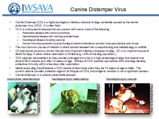
Canine distemper virus
(CDV),
belongs
to the genus
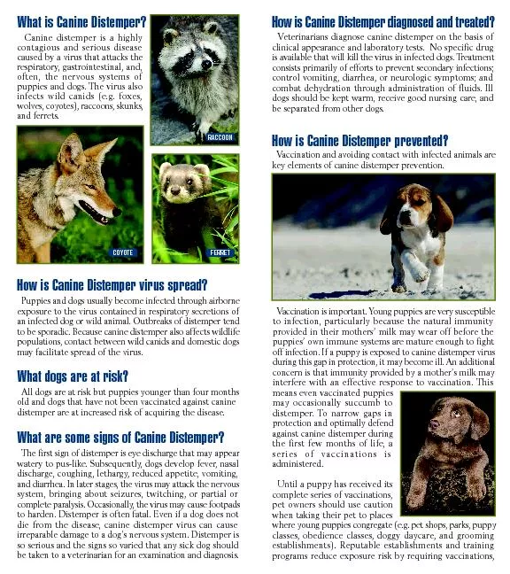
Morbillivirus (family, Paramyxoviridae).CDV is closely related to human measles virus and rinderpest virusDogs and other Canidae such as coyotes, foxes, and wolves; Procyonidae (raccoons, pandas); Mustelidae (ferrets, mink, skunks, otters). Large wild Felidae can also be affectedInfection of dogs can lead to a severe, multisystemic disease that primarily affects the gastrointestinal, respiratory, and neurologic systemsBrachiocephalic dogs have a lower prevalence than dolichocephalic breeds.World wide
ETIOLOGY
HOST AFFECTED
DISTRIBUTION
Slide3Fig: Anatomic sites
targeted by virus
Slide4Oronasal
contact with virus in
secretions or excretions, droplet nuclei, and large particle
aerosol transmission
MODE OF TRANSMISSION
PATHOGENESISVirusMonocytes within lymphoid tissue in the upper respiratory tract and tonsilsEntire RE SystemLymphatics and Blood
Slide5Viral
haemagglutinin
+Host Receptor (SLAM, Signalling lymphocytic activating molecules SLAM is expressed by immature
thymocytes
, activated lymphocytes, macrophages, and dendritic cells
Direct viral destruction of the lymphocyte population (CD4+ T cells), which occur within the blood, tonsils, thymus, spleen, lymph nodes, bone marrow, mucosa-associated lymphoid tissue, and hepatic Kupffer cells Initial Lymphopenia and Transient FeverSubsequently, 8-9 days post infection,there is a second stage of cell-associated viremia and feverAfter that it infects, respiratory, GIT, CNS, SKINS &additional Lymphoid cellsIn this stage, CDV infects a variety of cell lineage(Epithelial, mesenchymal, neuroendocrine, and hematopoietic cells, and forms intracytoplasmic but also
intranuclear inclusions)
Shedding of virus from all secretions and excretions at 5 days post infection, Before clinical
singns
Slide6CLINICAL SIGNS
The clinical signs depends on
:
virus strain, the age and immune status of
the host, as well as concurrent
infections with other viruses and bacteriaRespiratory form: fever, bilateral serous and nasal ocular discharges, conjunctivitis, and a non-productive cough and Secondary bacterial infection due to secondary bacterial infection lead to the development of mucopurulent nasal and ocular discharges and bacterial bronchopneumonia, with tachypnea, productive cough, lethargy, and decreased appetite.GI form: It lead to inappetence, vomiting, diarrhea, electrolyte abnormalities, and dehydration. Hyperkeratosis of nasal planumHyperkeratosis of foot pad
Slide7Nervous form:
the neurologic signs are often progressive due to neuronal necrosis and atrophy, and generally do not resolve, so dogs that recover often have residual neurologic deficits.
“Old dog encephalitis
” is a misnomer, poorly characterized progressive immune-mediated demyelinating leukoencephalomyelitis
, which occurs weeks to years after recovery from the acute infection and is difficult to diagnose
. A Myoclonus (involuntary twitching of isolated muscle groups) or chorea is common, when affected puppies are at rest. A “chewing gum” seizures, which localized to the head and jaw, with accompanying foamy hypersalivation.Other neurologic signs include obtundation, seizures, tremors, opisthotonos, tetraparesis, paraparesis, delayed placing reactions, ataxia, and, less commonly, behavioral abnormalities, compulsive pacing, and vestibular signs such as a head tilt, nystagmus, strabismus, and circlingOcular form : include blepharospasm, photophobia, uveitis, chorioretinitis, keratoconjunctivitis
sicca (KCS), keratitis, and
optic neuritis, which may be associated with blindness.
Slide8Enamel and dentin hypoplasia in recovered
animal.
Cutaneous form:
There is hyperkeratosis in the regions of footpad and nasal planum
epithelium.
Puppies with hyperkeratosis, also known as “hardpad,” have thickened, crusty footpads that resemble those of adult dogs.The most common secondary infections in distemper are secondary bacterial infections that contribute to bronchopneumonia is Bordetella bronchiseptica.The transplacental infections may cause h infertility, stillbirth, or abortion and with neurologic signs in puppies that are less than 4 to. 6 weeks of ageDogs that have recovered from CDV infection may develop enamel hypoplasia/chorea/keratoconjunctivitis Sicca/gold medallion lesions(healed chorioretinitis, a hyperreflective circular lesions).
Slide9DIAGNOSIS:
Clinical signs and
sypmtoms, especially myoclonus.
In dogs with
respiratory signs
, the presence of conjunctivitis, blepharospasm, and ocular discharge should raise suspicion for distemper. Distemper should always be considered as a diagnosis in young dogs with neurologic signsMicrobiologic Tests: This includeVirus Isolation, Cytologic Demonstration of CDV Inclusions, ELISA assays, Serology, RT-PCR.Enamel hypoplesia
Slide10TREATMENT and PROGNOSIS:
Dogs
with severe respiratory and gastrointestinal disease require:
IV fluids
Antimicrobial
drugs/antimicrobial drug combinations, such as ampicillin and a fluoroquinoloneAntiemeticsA single IV dose of dexamethasone or tapering anti-inflammatory doses of glucocorticoids has been advocated to halt or reduce the severity of progressive CNS signs.Oral vitamin A and C may be advocatedAnticonvulsants such as diazepam and, for chronic management, potassium bromide or phenobarbital can be used to treat seizures and prevent the spread of a seizure focus.Note:The prognosis for dogs with severe neurologic signs is poor.
Slide11Immunity and
Vaccination
Immunity to CDV requires antibodies as well as cell-mediated immunity
Disease can be prevented through immunization with attenuated live or recombinant vaccines
Attenuated live
vaccines should be given >than 6 weeks of ageFor dogs older than 16 weeks of age that are brought to the veterinarian for their first vaccination, two doses of vaccine should be given 3 to 4 weeks apart.Notes to be remember:Attenuated live CDV vaccine virus can cause encephalitis when administered to immunocompromised dogs and puppies less than 6 weeks of age.Postvaccinal encephalitis usually occurs 3 to 20 days after vaccinationassociated with certain batches of vaccines that contain the Rockborn strain.Attenuated live CDV vaccines to dams immediately after whelping has been associated with the development of encephalitis in the puppies