
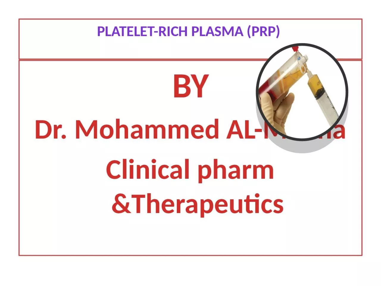
BY Dr Mohammed ALManna Clinical pharm ampTherapeutics The most natural injectable Plateletrich plasma PRP Definition Plateletrich plasma PRP is blood plasma that has been enriched with platelets ID: 930751
Download Presentation The PPT/PDF document "Platelet-rich plasma (PRP)" is the property of its rightful owner. Permission is granted to download and print the materials on this web site for personal, non-commercial use only, and to display it on your personal computer provided you do not modify the materials and that you retain all copyright notices contained in the materials. By downloading content from our website, you accept the terms of this agreement.


Slide1
Platelet-rich plasma (PRP)
BYDr. Mohammed AL-MannaClinical pharm &Therapeutics
Slide2The most natural injectable
Platelet-rich plasma (PRP)
Slide3Definition Platelet-rich plasma (PRP) is blood plasma that has been enriched with platelets. By PRP technique we will obtain 5-10 folds of platelets count ; about 2- 4.2million /mm3) PRP contains ,and releases (through degranulation) several different
growth factors and other cytokines that stimulate healing of bone and soft tissue.
Slide4Preparation of PRP
10-15 ml blood sample was aspirated and placed in sterile test tubes ( that contained 0.5 mL of 10% tri sodium citrate as the anticoagulant) and centrifuged in a standard laboratory centrifuge for 10 min at 4000 rpm. Subsequently, the yellow plasma (containing buffy coat with platelets and leukocytes) was taken up using a micropipette or syringe
Slide5THANK YOU
Slide6At the upper of test tube platelets and WBCs will be precipitated .This part should be taken and activated by mixing with thrombin , collagen, calcium chloride or calcium gluconate as an activator. 1 : 10 (vol /vol ) mixture. PRP and activator was incubated for 5 min at room temperature as become ready to be used.
Slide7Clinical applicationsThe platelets collected in PRP are activated by the addition of thrombin , collagen or
calcium , which induces the release of the growth factors and cytokines from alpha granules. The growth factors present in PRP include:
Slide8Vascular endothelial growth f
Slide9Clinical applicationsThe resulting PRP accelerates endothelial, epithelial, and epidermal regeneration. They stimulate angiogenesis, enhance collagen synthesis, promotes wound healing, decreased dermal scarring, enhanced
hemostatic response and reverse the inhibition of wound healing caused by glucocorticoids. High leukocyte concentration in PRP has an added antimicrobial effect.
Slide10THANK YOU
Slide11Injection levels are both in dermis and hypodermis,,
Slide12indications of PRP:1 : Sport related injuries:Achilles tendinitis,,tennis elbow... muscle injuries bone repair
regeneration of nerve injuryClinical applications
Slide13Clinical applications2.osteoarthritisThe use of PRP to stimulate cartilage repair is a particularly exciting application. It is thought that PRP can stimulate chondral anabolism, reduce catabolic processes, and may improve overall joint homeostasis reducing synovial membrane hyperplasia .
Slide14Clinical applicationsIt has been demonstrated that PRP administered three times weekly to patients with grade 3 or 4 knee OA reported improvements in their quality of life, and reduced levels of pain, and had increased cartilage thickness at the 6-month follow up.
Slide15Clinical applications3.Cardiac muscle surgeryRecent tissue engineering researches suggest that cells and PRP-derived growth factors together into biomaterials have opened new horizons in the treatment of myocardial infarction and other heart diseases.
Slide16Clinical applications4.Dermatological diseasesA.
Strech marks ((Striae))
Slide17Clinical applications
B. Postacne and chicken box scar – PRP combined with centrifuged fat tissue , fractional laser resurfacing , dermal roller or derma pen improve cosmetic appearance of scars.
Slide18After PRP
Before PRP
Slide19Clinical applicationsc. Hair lossHair loss
disorders – PRP has been shown to reinvigorate dormant hair follicles and stimulate new hair growth .FGF prolonged anagenVEGF and PDGF proangiogenic
Slide20Where is my hair?!
Slide21Type of hair lossPRP can be used in different type of hair loss such as Alopecia androgenticaAlopecia areata Telogen effluvium
Slide22Clinical applications5. Cosmatics
Facial wrinklesFace liftingTear trophDark circleFacial rejuvenation – PRP injections can treat wrinkles, photodamage and discoloration in conjunction together with other treatment modalities
Slide23Clinical applications
Slide24THANK YOU
Slide25Indications
as a whole
Slide26Clinical applications
Slide27Clinical applications6. ulcersVenous and arterial leg ulcers
Diabetic foot ulcersPressure ulcers (bedsores)Skin graft donor sitesFirst and second degree thermal burnsSuperficial injuries, cuts, abrasions and surgical wounds
Slide28THANK YOU
Slide297. Dental usesPlatelets Rich Plasma is a new approach to tissue regeneration and it is becoming a valuable adjunct to promote healing
in many procedures in dental and oral surgery.The use of PRP in surgical practice could have beneficial outcomes, reducing bleeding and enhancing soft tissue healing and bone regeneration. Since PRP is free from potential risks for patients, not difficult to obtain and use, it can be employed as a valid adjunct in many procedures in oral and dental surgery. *Studies conducted on humans have yielded promising results regarding the application of PRP to many dental and oral surgical procedures (i.e. Jaw osteonecrosis ,,, tooth extractions,,, periodontal surgery,,, implant surgery).
Slide30Clinical applicationsA) Jaw osteonecrosis: Bisphosphonates (BPs) are a class of drugs commonly used to treat bone metastasis and various bone diseases such as osteoporosis). Bisphosphonate-related osteonecrosis of the jaw (BRONJ) is a common complication in patients who received BPs, especially
intravenously. The use of PRP has also been proposed in the management of bisphosphonate -related osteonecrosis of the jaw (BRONJ) with the aim of enhancing wound healing and bone maturation. The combination of necrotic bone curettage , antibiotics and PRP application seem to be encouraging for the treatment of refractory jaw osteonecrosis, as it has proven successful outcomes with minimal invasivity.
Slide31
Slide322) Alveolar socket:Alveolar osteitis is inflammation of the alveolar
bone. Classically, this occurs as a postoperative complication of tooth extraction.Alveolar osteitis usually occurs where the blood clot fails to form or is lost from the socket (i.e., the defect left in the gum when a tooth is taken out). This leaves an empty socket where bone is exposed to the oral cavity, causing a localized alveolar osteitis .This specific type of alveolar osteitis is also known as dry socket or, less commonly, fibrinolytic alveolitis, and is associated with increased pain and delayed healing time.Dry socket occurs in about 0.5–5% of routine dental extractions , and in about 25–30% of extractions of impacted mandibular third molars (wisdom teeth which are buried in the bone)
Slide33Alveolar socket:Factors considered to be indicated for PRP ”risk factors” include:Age,SexPoor oral hygiene Immunosuppression and diabetes
smoking status,concurrent use of oral contraceptive use,presence of oral clenching/grinding habits (bruxism),Impacted tooth
Slide34Alveolar osteitis (AO) is characterized by severe throbbing pain that begins within 3 to 5 postoperative days and is usually refractory to NSAIDs and narcotic analgesics.
RxPatients receiving PRP treatment immediately following tooth extraction significantly reduced the incidence of AO by 62% .*PRP may be of benefit because it helps initiate clot formation, provides growth factors to facilitate the healing process, and contains concentrated white blood cells to inhibit infection.
Slide35procedure:Blood samples of approximately 10 mL each were aspirated and placed in 2 sterile test tubes .Tubes centrifuged for 10 minutes centrifugation separated the whole blood into 3 layers: red blood cells (RBCs) on the bottom, platelet-poor plasma (PPP) on the top, and between these two layers, a small band of PRP.
The PRP layers were aspirated by 3ml syringe and were then placed into an empty tube (not containing trisodium citrate) and cetrifuged . The next step was to coagulate, or gelling the PRP by 0.05 mL (5 units ; 3drops) of 10% calcium chloride.
Slide36procedure:. This step leading to the formation of fibrin clot that is rich in platelets. This gelling process usually takes between 10 and 20 minutes.The PRP was dropped into a sterile surgical stainless steel cup.
The extraction socket was prepared by curetting and irrigating with 10 to 20 mL of sterile saline.The gelled PRP fibrin clot was removed from the sterile surgical cup with sterile pliers and placed into the prepared extraction socket. Sutures were then placed to achieve primary closure and to help retain the PRP clot.
Slide37Clinical applications3) Periodontal surgeryTechnique of application in periodontal intrabony defects
(bone loss) . The periodontal defect site is opened and debrided. The PRP gel is placed into the defect site and sutured . *PRP can be mixed with a graft material (bone graft) and placed in the defect . PRP results in early consolidation and take up of the graft. All these treatment modalities have shown good clinical results. Major Benefits of PRP “ osteogenesis by releasing growth factors at the local site. Use of PRP has shown to improve the rate of bone formation by 1.62 to 2.18 times that of the controls Note : There has been reported in the literature a 15% to 30% improvement in trabecular bone density when platelet rich plasma factor is added to the graft
Slide38Clinical applications4. Dental implant surgery : the American Academy of Implant Dentistry demonstrate that
PRP therapy can accelerate bone and tissue growth and wound healing and help assure long-term success of dental implant placements.PRP treatments can jump start bone growth and implant adherence in just two weeks, which cuts down the time between implant placement and affixing the permanent crown.PRP triggers rapid growth of new bone and soft tissue. "There is very little risk because we are accelerating the natural process in which the body heals itself. For dental surgery applications PRP is mixed as a gel that can be applied directly in tooth sockets and other sites. It also is effective in cases when bone grafts are required to foster proper bone integration for implants. Growth factors in PRP preparations help the grafts bond faster with the patient's own bone.
Slide39Clinical applicationsBMP (Bone Morphogenic Protein) is one of the growth factors that is released with PRP placement and it has been shown to stimulate the formation of new bone in humans
Slide40SIDE EFFECTS OF PRPMild Pain and headache for first 12-24 hours.Swelling and redness for 1-3 days .
bruising (2 weeks,, masked by make-up,)In very rare cases, skin cellulitis may occur which can be treated with antibiotics and cold compresses
Slide41PRP in people over 60 years of ageAlthough not impossible but they are poor responders.
Slide42Contraindication for PRP Active skin
disease (such as : SLE, porphyria, )Cancer chemotherapy.Hypertrophic diseases (scars) and angiogenesis –based disease (psoriasis)Severe metabolic and systemic disorders (Uncontrolled diabetes and severe asthma )Abnormal platelet function. NSAIDS during one week beforeHistory of recent stroke
(heavy)
Cigarette
smoking
Active infection
at the injected Site
Anti-coagulation therapy
Slide43What to avoid post—op care Avoid massaging the treated area for first 48 hours after the treatment
. Also avoid facial peels , intralesional steroid injection and laser treatments for the first 4 weeks.
Slide44THANK YOU