

i 3EM Thursday 6 th July 2017 Introduction Hand injuries account for between 510 of attendances in emergency departments EDs in the UK 2030 are improperly diagnosed and go unrecognised ID: 933277
Download Presentation The PPT/PDF document "Finger injuries Dr Alex Novak" is the property of its rightful owner. Permission is granted to download and print the materials on this web site for personal, non-commercial use only, and to display it on your personal computer provided you do not modify the materials and that you retain all copyright notices contained in the materials. By downloading content from our website, you accept the terms of this agreement.


Slide1
Finger injuries
Dr Alex Novak
i
3EM Thursday 6
th
July 2017
Slide2Introduction
Hand injuries account for between 5-10% of attendances in emergency departments (EDs) in the
UK - 20-30
%
are
improperly diagnosed and go
unrecognised
The
hand is the most commonly injured part of the
body
The
fingertip the most common hand injury
Challenge to distinguish
an occult tendon, ligament or nerve injury from the uncomplicated laceration or crush injury
Important
to identify who is suitable for ED management vs specialist referral
Slide3Tendons (FDS and FDP)
The FDS and the FDP tendons travel distally from the forearm through the carpal tunnel, after which they traverse a fibro-osseous tunnel in each digit to insert in the respective
phalanges
The
profundus
tendon pierces that of the
superficialis
over the proximal
phalanx
The metacarpal heads, phalanges and intervening joints
= dorsal wall
annular pulley system and fibrous flexor sheath
=
anterolateral wall
The fibrous sheaths are lined by the synovial membrane, which reflects around each
tendon
Slide4FDS and FDP
Slide5Nerves
ULNAR NERVE:
Ulnar 2
FDP tendons to the little and ring fingers; the other long finger flexors are supplied by the median
nerve
Sensation
over the ulnar side of the hand and little
finger
Dorsal
ulnar region of the hand via the dorsal cutaneous branch of the ulnar
nerve
MEDIAN NERVE:
sensation
over the palmar index, middle fingers, thumb, and proximal palm near the
thenar
eminence
Test motor function with
abductor
pollicis
brevis action, thumb abduction with palm up, raising the thumb to
perpendicular
Weakness or absence of flexion of the IPJ of the thumb (FPL) and the DIPJ of the index finger (FDP) against resistance, if present, are due to a more proximal lesion (
anterior interosseous nerve
).
Slide6Finger innervation
Slide7Assessment - History
Age
Hand dominance
Occupation/hobbies
Where, when and how did the injury occur?
What was the position of the hand at the time of
injury?
Both
hands should be compared to better assess baseline function
Slide8Finger cascade
Slide9Examination - general
deformity
, open wounds, bruising and
swelling
Pallor
or cyanosis
- vascular
compromise
Loss
of cascade may indicate a flexor tendon
injury
In
small children, uncooperative or
obtunded
patients, in addition to hand posture, tendon continuity can be assessed by squeezing the forearm muscles while observing the
fingers
With
intact extensors, passive wrist flexion causes finger
extension
When
flexor tendons are intact wrist extension leads to flexion of the
fingers
Slide10Nerve Injury
A
bsence
of sweating =
sign of nerve injury due to loss of sympathetic
innervation
Two-point
discrimination
– static (6mm)
or
dynamic (4mm)
Sensory
loss following a proximal crush injury or closed fracture suggests ongoing nerve compression and may require surgical
decompression
Sensory loss in relation to a hand laceration is a sign of nerve division and requires surgical
exploration
Temporary nerve malfunction may occur in a closed injury due to mechanical trauma (
neuropraxia
)
– use serial
examinations by the same
observer
Slide11Sheaths and Pulleys
Sheaths
of the thumb and little finger extend proximally into the palm as the radial and ulnar bursae
respectively
-
extend below the flexor retinaculum and communicate in about 50% of patients
The
annular and cruciform pulleys preventing bowstringing when flexing the metacarpophalangeal (MCP), proximal interphalangeal (PIP), and distal interphalangeal (DIP) joints
Three cruciform pulleys (C1-C3) and five annular pulleys (A1-A5) exist
From a biomechanical advantage point the A2 and A4 pulleys are considered the most important to prevent
bowstringing
Slide12Sheaths and Pulleys
Slide13Flexor Tendon Injury Zones
Slide14Flexor Tendon Injury Zones
A distal-to-proximal 5-zone (I-V) classification system has been developed based on location, treatment considerations and prognosis
I -
Zone
I
contains only the FDP tendon and extends from the insertion of the FDP to the insertion of the FDS tendon.
II -
Zone
II
is the area extending from the insertion of the FDS tendon to the distal palmar crease (proximal end of the A1 pulley). This area is also known as 'No-Man's land', due to the shared flexor sheath and a higher risk of adhesions.
III -
Zone
III
is the palm area from the distal palmar crease (proximal end of the A1 pulley) to the distal border of the transverse carpal ligament.
IV -
Zone
IV
is within the carpal tunnel.
V -
Zone
V
is proximal to the carpal tunnel in the distal forearm
.
Slide15Thumb flexor tendon injury zones
Thumb flexor tendon injury zones differ from the fingers as the thumb has one less
phalanx
TI
-
Zone TI
is from the insertion of the flexor
pollicis
longus (FPL) to the proximal part of the A2 pulley.
TII -
Zone TII
is from the proximal part of the A2 pulley to the distal part of the A1 pulley.
TIII -
Zone TIII
is proximal to the A1 pulley as far as the carpal tunnel.
Slide16Finger Extension
Combination
of extrinsic and intrinsic muscle
action
E
xtrinsic
extensors =
primarily responsible for MCPJ extension, with extension of the IPJs being primarily an intrinsic
function
The long extensors of the fingers are the extensor
digitorum
communis
(EDC), reinforced by the extensor
indicis
and the extensor
digiti
minimi
, joining the appropriate tendons of EDC on the ulnar side
Slide17Slide18Finger extension (2)
As the tendons pass over the MCP joints they are stabilised by tough transverse fibres called sagittal bands
The tendons of the EDC terminate in each finger as an aponeurotic extensor expansion, covering the dorsum of the proximal phalanx and the side of its base.
Attaches by a central slip into the base of each middle phalanx, and by two lateral slips to the base of each distal phalanx
Slide19Extensor Tendon Injury Zones
Slide20Extensor Tendon Injury Zones
I - area
over the DIP joint and distal phalanx. Disruption of the tendon will cause mallet
finger/swan-neck deformity
II - over
the middle phalanx; assessment and treatment are the same as for zone I
injuries
III - over
the PIP joint. Injury here can result in a boutonnière's
deformity
IV
- on
the proximal phalanx
-treated
like zone III
injuries
V - over
the MCP
joint
VI - dorsum
of the hand. The tendons are very superficial here and can be easily
damaged
VII
injuries
- wrist
and multiple tendons; these should be evaluated by a hand
surgeon
VIII
injuries
-
in the distal forearm. Injuries in this location often require tendon retrieval for complete lacerations and may need to be performed in the operating
room
Slide21Slide22Flexor Tendon Injuries - Tendon Evaluation Tests
Testing the flexor
digitorum
superficialis
(FDS)
The patient should bend the finger whilst the others are held in full extension (thereby inactivating the deep flexors). The DIPJ should be flaccid. The exception is the index finger, which has a separate muscle belly so that extending the other digits does not isolate the FDS.
For FDS to the index finger – test by checking the resisted PIPJ flexion while keeping the DIPJ extended.
Slide23FDP test
Testing the flexor
digitorum
profundus
(FDP)
With the examiner holding the PIPJ in extension, the patient should be asked to flex the tip of the finger
.
Slide24Extensor tendon test
Testing the extensor tendons
The fingers should be straightened against resistance. The long extensors straighten at the MCPJ, and resistance should be applied to the dorsum of the proximal phalanx.
Extension at the PIPJ can be caused by the intrinsic muscles. Observe for loss of active extension at the DIPJ, i.e. a mallet deformity
.
Slide25FPL test
Testing the flexor
pollicis
longus (FPL)
Hold the thumb over the proximal phalanx and ask the patient to bend the
tip
Slide26EPL test
Testing the extensor
pollicis
longus (EPL)
With the patient's hand palm-down on a table, ask the patient to lift up his/her thumb, against resistance
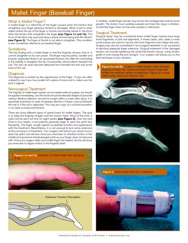
Slide27Mallet Finger
Slide28Extensor Tendon Injuries - Mallet Finger (Zones I and II)
Zone
I and Zone II injuries may result in a mallet deformity, due to loss of continuity of the conjoined lateral bands at the DIP joint
Usually
a direct blow that forcibly flexes an extended
finger
If
left untreated, apart from being painful, the digit becomes
hooked/swan-neck
deformity
(compensatory
hyperextension
@
PIPJ)
Open
injuries
- ref to hand
surgeon for primary
repair
Closed
mallet finger injuries
-
treated conservatively
May be
due to bone or soft-tissue injury
– need XRs
Slide29Mallet finger- treatment
Conservative
treatment
- continuous
splinting of the DIPJ in neutral or slight hyperextension for at least six
weeks
A
well-fitting splint for a mallet finger
=
vital to ensure compliance and avoid skin breakdown, the main complication of conservative
treatment
PIPJ
must be left free to allow mobilisation and prevent
stiffness
Refer if:
The absence of full passive extension (indicating possible bony or soft-tissue entrapment requiring surgical intervention)
Joint subluxation or an avulsion fracture of more than one-third of the articular surface
Slide30Extensor Tendon Injuries - Rupture or Division of the Central Slip
Slide31Extensor Tendon Injuries - Rupture or Division of the Central
Slip (1)
Zone
III injuries may involve rupture or division of the central slip
Axial
loading or forced flexion with the PIPJ in
extension, or
volar dislocation of the PIPJ
Variable presentations–
e.g. acute boutonniere deformity,
volar
dislocation or painful swollen PIPJ
M
aximal
localised tenderness over the dorsal aspect of the PIPJ, at the insertion of the central
slip +/- bruising
Active
extension at the PIPJ
does not exclude a rupture
as full
extension may still be achieved by the lateral
bands
Closed
rupture of the central slip over the PIPJ is easily
missed
Slide32Extensor Tendon Injuries - Rupture or Division of the Central Slip
(2)
Elson's
test - PIP
joint of the injured finger flexed 90° over the edge of a
table. Patient
then tries to extend the PIP joint of the injured finger against resistance. The absence of extension force at the PIP joint and fixed extension at the DIP joint are signs of complete rupture of the central
slip
will
not demonstrate a partial rupture, and may be limited by
pain
X-ray may show an avulsion
fracture
If
a central slip rupture is known or strongly suspected, the PIPJ should be splinted in a static extension splint, leaving the DIPJ
free
Further follow-up in a hand clinic is
required
Slide33Nerve Injury
The early recognition of nerve injury is important as primary repair has been found to be superior to delayed repair
Results of digital nerve repair are variable; in a review of 109 cases, no patients regained normal sensation, although 83% did achieve sensory results that could be classed as 'good
'
Results are better in children than in adults
Loss of motor/sensory function
Nerve
injury is also suggested by dry, shiny skin that does not wrinkle when immersed in
water
– this is due to a loss of sympathetic
innervation
The tactile adherence test
- loss
of friction in the
denervated
area due to absent
sweating
Slide34Extensor
Tendon Injuries -
Lacerations
(Zone IV)
Zone IV tendon injuries over the proximal phalanx are usually due to lacerations.
The ED management should include a wound exploration under local anaesthetic cover.
Confirmed tendon injuries need referral to a hand surgeon for tendon repair, splinting and follow-up.
Slide35Fingertip injuries
= most
common hand injuries in
adults and children
Damage
to the nail bed is reported to occur in 15-24% of fingertip injuries
Defined
as any soft tissue, nail or bony injury distal to the dorsal and volar skin creases of the distal interphalangeal joint and insertions of the long flexor tendons of the fingers or
thumb
Injury
can include damage to skin and soft tissue, bone (distal phalanx) or to the nail and nail
bed
Approximately
half of fingertip injuries have an associated fracture of the digit, usually the distal tuft of the terminal
phalanx
Fractures proximal to the waist of the terminal phalanx or intra-articular fractures require specialist surgical assessment to determine the need for internal
stabilisation
Slide36Fingertip Injuries - Fingertip Anatomy
Slide37Fingertip Injuries - Fingertip Anatomy
The fingernail and its underlying matrix are supported by more than half the bone length of the underlying terminal
phalanx
In the
distal pulp of the finger the skin is stabilised by fascial bands running from skin to bone, which contain
loculated
fat; this arrangement stabilises the fingertip pulp skin under
pressure
Nail
growth is slow, approximately 0.1 mm
daily,
but often with an initial delay following traumatic avulsion, hence may take 4-6 months to grow back fully and up to 12 months to achieve
maturity
Slide38Fingertip Injuries - Fingertip Amputations
Type I
Fingertip soft-tissue loss only
Type II
Fingertip loss at the level of the proximal third of the nail plate
Type III
Fingertip loss at the level of the
eponychial
fold
Type IV
Fingertip loss proximal to the DIP
joint
Slide39Fingertip Injuries - Fingertip Amputations
Oblique
fingertip defects
Volar
Dorsal
Lateral
Slide40Fingertip injuries suitable for management by an ED specialist
Superficial
skin loss defect <1 cm
2
in Zone I
Transverse Type I fingertip amputations
Type I and Type II fingertip amputations in children
Oblique partial fingertip amputation without bone exposure
Stable fracture needing
splintage
only
NB 1: Follow-up
clinic and dressings facilities, as well as access to a physiotherapist, are
required
NB 2: children
have greater tissue regeneration and modelling capacity.
Slide41ED Treatment of Fingertip Injuries
Local
anaesthesia ring
block (essential
for assessment, treatment and
analgesia)
Cleanse thoroughly, remove dirt and foreign material
Trim off any devitalised tissue
Non-adherent dressings to be changed and the wound inspected 2-3 times weekly
A stable fracture of the terminal phalanx may be externally splinted for 2-3 weeks
A 1 cm
2
defect takes an average of five weeks to
heal
Fingernail
and its underlying matrix are supported by more than half the bone length of the underlying terminal
phalanx
Type
I and Type II amputations have sufficient bone support for straight nail regrowth without hook nail
deformity
Avulsed
nail can be placed in the nail fold, which acts as a template and stent for the nail bed and also decreases discomfort by acting as a natural protective cover
Slide42Fingertip injuries NOT suitable
for management by an ED specialist
Type
II or higher partial amputation of the fingertip
Oblique fingertip partial amputation with bone exposure or fracture
A large soft-tissue defect >1 cm
2
An unstable fracture of the terminal phalanx
Slide43Preparation
a patient for transfer to a hand specialist
unit
IV fluids
if the patient needs fasting for a general anaesthetic and for patient hydration
IV
antibiotics e.g.
first-generation cephalosporin
Control pain
with IV morphine or a digital block
Get a radiograph
of the digit and also of the amputated segment (if available)
Clean and dress the finger stump
with non-adherent dressing, and wrap the stump lightly with sterile dressings and bandage
Elevate
the affected hand in a
sling
Slide44Care of the amputated part
Remove
any foreign material
from the exposed soft tissues
Clean
the amputated part with saline, and
wrap
it in saline moistened gauze (damp, not soaking wet)
Place the wrapped segment in a
plastic bag
Place the bag into a
container filled with ice mixed with
saline
DO
NOT place the amputated part directly on
ice
Slide45Subungual Haematoma
Slide46Subungual Haematoma
Crush injuries of lesser severity may cause subungual
haematoma
Small
nail bed laceration with resultant bleeding occurs, which cannot drain from an intact
nail
Painful
subungual haematomas should be drained
for relief
P
rospective
studies comparing simple haematoma decompression versus nail plate removal and formal nail bed repair have shown no notable difference in
outcome
Therefore if
the nail plate is still adherent to the nail bed and not displaced out of the nail fold, regardless of the size of the subungual haematoma, conservative treatment is
recommended
= Needle
trephining using a hot paper clip or battery powered cautery
Slide47Nailbed lacerations
Slide48Nail bed lacerations (1)
Simple
or stellate nail bed lacerations without underlying fractures have a better prognosis than nail bed avulsions
Can
be repaired by separating and removing the nail plate from the nail bed with fine
scissors
N
ail
bed is carefully repaired with fine absorbable sutures
The
removed nail is trimmed of sharp edges and replaced in the nail fold to act as a stent for the nail bed, a template for the new nail, and as a protective cover to reduce pain and
discomfort
A
transverse suture through the nail and lateral folds will help retain the nail in
position
R
etain
the nail for four weeks before discarding it by cutting the retaining
sutures
Slide49Nail bed lacerations with associated fractures
N
ail
bed =
supported internally by the terminal phalanx bone and externally by the nail
plate
If laceration =
associated with stable
undisplaced
fractures of the terminal phalanx bone,
then simple
nail bed
repair/external splint
If displaced
or unstable fractures
then refer
for specialist treatment
as can
result in nail bed irregularity, scarring, nail plate detachment and late nail
deformity
Specialist
referral is required due to a high risk of hook nail deformity in: nail bed lacerations with an underlying displaced or unstable fracture; nail bed avulsion; type III and IV fingertip injuries.
Nail
bed avulsions
- refer
to a hand specialist as meticulous repair (e.g. loupe magnification, nail bed grafting or rotational flap surgery) may be
required
Slide50The Thumb - movement
Eight muscles are responsible for thumb movement, and these may be divided into long (from the forearm) and intrinsic hand.
FPL
is the only long flexor of the thumb, inserting into the base of the distal phalanx, and flexing the IPJ.
EPL inserts into the distal phalanx, extending the IPJ.
EPB inserts into the proximal phalanx, extending the MCPJ.
AbPL
inserts into the first metacarpal, and carries the thumb laterally from the palm.
The
AdP
, FPB and
AbPB
all insert into the base of the proximal phalanx.
The FPB,
AbPB
and OP make up the
thenar
eminence and are supplied by the median nerve.
Slide51Thumb MCP joint stability
Ulnar side
stability -
static and dynamic
mechanisms
Static
stability
- main
and accessory UCLs, the volar plate,
dorsal capsule
dynamic
stability - adductor
pollicis
muscle and tendon in the first web space of the
hand
UCL proper - from the head of the thumb metacarpal bone to the volar aspect of the proximal phalanx
Tightens in flexion and relaxes in extension of the MCP joint
Accessory UCL lies anterior to the UCL proper and inserts into the volar plate
Tightens in MCP joint extension and relaxes in flexion
Adductor
pollicis
aponeurosis inserts to the dorsal thumb tendons and capsule of the MCP joint
Slide52Ulnar Collateral Ligament
Slide53Ulnar Collateral Ligament Injury
(Gamekeeper’s/Skier’s thumb)
UCL
injuries may also involve injuries to the accessory UCL, volar plate, dorsal capsule, adductor
pollicis
insertion, and
associated
fractures of the proximal
phalanx
Ultrasound has been shown to improve the positive predictive value of clinical examination alone from 80% to 94% in a study undertaken in a British ED.
Other options include stress radiography and arthrography.
Usually conservative treatment - immobilisation
in a short thumb
spica
cast for four weeks to allow the ligament to heal. Thereafter, the cast can be discarded in favour of a short thermoplastic splint.
Supervised mobilisation is commenced at four weeks with all splints discarded after six weeks
Patients should avoid stressful activities with the injured thumb for 10-12 weeks
Slide54Slide55Slide56UCL injury - indications for referral to a hand clinic
Stener
lesion
Complete rupture of the UCL
Displaced, rotated or large fracture fragment of the base of the proximal phalanx bone
Subluxation or instability of the MCP joint
Ongoing uncertainty of the severity of the UCL rupture or
Stener
lesion
Slide57Thumb spica
Slide58Slide59Volar plate injury
Slide60Buddy strapping
Slide61PIPJ dislocation
Slide62PIPJ dislocation - reduction
Slide63Phalangeal fractures
Unlike distal phalanx fractures, proximal and middle phalanx fractures require precise alignment for good functional and cosmetic
outcomes
M
ajority
of pharyngeal fractures
=
undisplaced
transverse
fractures
– Rx
“
buddy-strapping”
Unstable pharyngeal fractures include oblique fractures,
malrotated
fractures, and angulated
fractures -
manipulation under digital block to reduce to adequate
alignment
Once
alignment has been
achieved,
the fracture should be splinted in extension and refereed to the outpatient hand clinic
Slide64Ring block
Slide65Ring Block
Slide66Summary
Finger injuries are very common
Surprisingly complex – apparently small injury can result in significant loss of function
Understanding of anatomy crucial to accurate diagnosis and appropriate management
Slide67References
Acute
Finger Injuries: Part I. Tendons and Ligaments
CHRISTIAN
J. MEKO
Am Fam Physician.
2006 Mar 1;73(5):810-816.
RCEM learning
LITFL