
Its size same the nucleus of lymphocyte and has diameters of 6 to 85 μm RBC in Healthy person Normocytic size is normal Normochromic Hb content ie colour is normal ID: 935936
Download Presentation The PPT/PDF document "Red cell morphology: In healthy person,..." is the property of its rightful owner. Permission is granted to download and print the materials on this web site for personal, non-commercial use only, and to display it on your personal computer provided you do not modify the materials and that you retain all copyright notices contained in the materials. By downloading content from our website, you accept the terms of this agreement.


Slide1
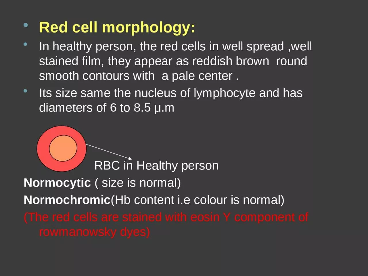
Red cell morphology: In healthy person, the red cells in well spread ,well stained film, they appear as reddish brown round smooth contours with a pale center .Its size same the nucleus of lymphocyte and has diameters of 6 to 8.5 μ.m RBC in Healthy personNormocytic ( size is normal)Normochromic(Hb content i.e colour is normal)(The red cells are stained with eosin Y component of rowmanowsky dyes)
Slide2Variation in RBCs morphology due to:1-Abnormal erythropoiesis :production of RBC only2- increased erythropoiesis3-decreased Hb formation4-RBC damageThese causes result in the following variation:Anisocytosis → variation in size (micro or macro) Poikilocytosis → variation in shapeVariation in colourVariation in content
Slide31. Variation in Size: Normal
Liver disease
Alcoholism
Megaloblastic
anaemia
Macrocyte
(Large) Iron deficiency anemia. Haemoglobinopathy.Microcyte (small)
Slide42-Variation in shape: Extra medullary haemopoiesis
Poikilocyte (tear shape)
Hereditary elleptocytosis
Elliptocyte (rod like)
Ovalocyte (oval)
DIC → Disseminated intravascular coagulation. Burns Cardiac valves disease
Shistocyte
(RBC fragments,
(
pyknocyte
, helmet cell ,bite cell)
Sickle cell anaemia
Sickled cell (Banana shape)
Slide5Burr cells (Spine projection)
liver disease
renal failure
Crenation ((acanthocyte)) or spur cells
liver disease
renal failure
Ecchinocyte
- Iron deficiency anaemia Pencil cell
G6pD deficiency anaemia
Basket cell
G6PD deficiency anaemia.
Blister cell
Slide63- Variation in colour:(colour paler)Than normal
1. Hypochromasia
Dimorphic picture
non-uniform color of erythrocyte
2. Anisochromasia
iron deficiency
anaemia liver diseasethalassemiapost splenectomy3. Target cells
Very thin cells with
colour
less central part.(ring shape)
4.
Leptocyte
Sphere like with deep colour and no central pallor found in hereditary spherocytosis.
5. Spherocyte Pale greenish – blue color.
6. Polychromasia
Cause by:
liver disease
alcoholism
7. Stomatocyte