
oligodendrocytes in the active areas of a chronic active MS lesion Staining for IFNa b e h k in green and in a for B cells CD19 in d T cells CD3 and in g astrocytes GFAP and in j ID: 931722
Download Presentation The PPT/PDF document "Figure e-1 A: IFN-a is not expressed in ..." is the property of its rightful owner. Permission is granted to download and print the materials on this web site for personal, non-commercial use only, and to display it on your personal computer provided you do not modify the materials and that you retain all copyright notices contained in the materials. By downloading content from our website, you accept the terms of this agreement.


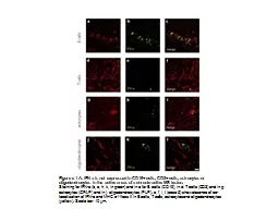
Slide1
Figure e-1 A: IFN-a is not expressed in CD19+ cells, CD3+ cells, astrocytes or
oligodendrocytes
in the active areas of a chronic active MS lesion.
Staining for IFN-a (b, e, h, k, in green) and in a for B cells (CD19), in d: T cells (CD3) and in g: astrocytes (GFAP) and in j:
oligodendrocytes
(PLP ). c, f,
i
, l (case 5) show absence of co-localization of IFN-a and MHC c
1
1
lass II in B cells, T cells, astrocytes and
oligodendrocytes
(yellow). Scale bar: 10 µm.
Slide2Figure
e-1B
:
Plasmacytoid
dendritic cells are not present in MS lesions.
a-c
:
Immunohistochemical
staining of formalin-fixed sections from MS
autoptic
specimens using
plasmacytoid
dendritic cell (
pDC
) markers as indicated.
d-f
:
Immunohistochemical
staining of formalin-fixed sections from a reactive lymph node using
pDC
markers as indicated. Note that CD123 also stains high endothelial
venules
. Scale bar: 200 µm.
g
:
Immunohistochemical
staining of BDCA2 in formalin-fixed normal brain. Scale bar: 100 µm.