
1Resolution restoration to histologic and functional normalcy Neutralization or removal of the various chemical mediator s N ormalization of vascular permeability ID: 933602
Download Presentation The PPT/PDF document "Outcomes of Acute Inflammation:" is the property of its rightful owner. Permission is granted to download and print the materials on this web site for personal, non-commercial use only, and to display it on your personal computer provided you do not modify the materials and that you retain all copyright notices contained in the materials. By downloading content from our website, you accept the terms of this agreement.


Slide1
Outcomes of Acute
Inflammation:
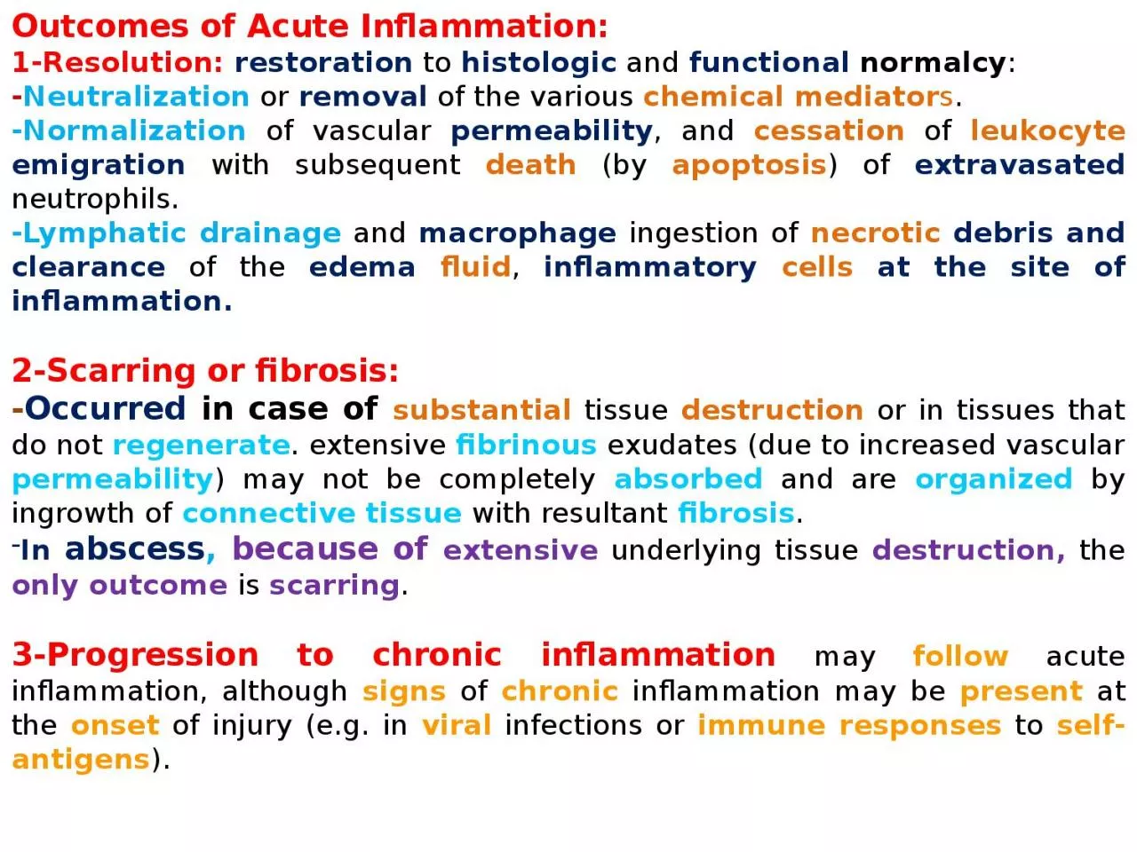
1-Resolution:
restoration
to
histologic
and
functional
normalcy
:
-
Neutralization
or
removal
of the various
chemical mediator
s
.
-
N
ormalization
of vascular
permeability
, and
cessation
of
leukocyte
emigration
with subsequent
death
(by
apoptosis
) of
extravasated
neutrophils
.
-Lymphatic drainage
and
macrophage
ingestion of
necrotic
debris and clearance
of the
edema
fluid
,
inflammatory
cells
at the site of inflammation.
2-Scarring or fibrosis:
-
Occurred
in case of
substantial
tissue
destruction
or in tissues that do not
regenerate
. extensive
fibrinous
exudates (due to increased vascular
permeability
) may not be completely
absorbed
and are
organized
by
ingrowth
of
connective tissue
with resultant
fibrosis
.
In
abscess
,
because of
extensive
underlying tissue
destruction,
the
only outcome
is
scarring
.
3-Progression to chronic inflammation
may
follow
acute inflammation, although
signs
of
chronic
inflammation may be
present
at the
onset
of injury (e.g. in
viral
infections or
immune responses
to
self-antigens
).
CHRONIC INFLAMMATION
prolonged duration
(
weeks
to
months
to
years
) in which
active
inflammation, tissue
injury
, and
healing
proceed simultaneously.
influx
of
lymphocytes
and
macrophages
with associated
vascular proliferation
and
scarring
.
Slide2Cauases
of chronic inflammation:
1-Viral infections.
2-Persistent microbial infections.
-
Mycobacteria
(
tubercle bacilli
),
Treponema
pallidum
(causative organism of
syphilis
), and certain
fungi
.
evoke
an immune response called
delayed hypersensitivity (
granulomatous
reaction)
.
3-Prolonged exposure to potentially toxic agents
.
-Exogenous
nondegradable
material such as
Inhaled
particulate silica
, which can induce (
silicosis
) in the
lungs.
-Endogenous
agents such as
chronically elevated plasma lipid components
, which may contribute to
atherosclerosis
.
4-Autoimmune diseases.
-
Immune
response to
self-antigens
and
tissues
. (e.g.,
rheumatoid arthritis
or multiple
sclerosis
).
Chronic inflammatory Cells and Mediators
1-Macrophages
-
(
mononuclear phagocyte system) in different organs.
-Filters
for
particulate matter
,
microbes
, and
senescent
cells.
-Sentinels
to alert the specific
components
of the immune system (
T and B
lymphocytes) to
injurious stimuli.
-Emigrate
to the site of injury within the
first 24 to 48
hours after
onset
of
acute
inflammation.
-Activated
by bacterial
endotoxin
,
cytokines
secreted by
sensitized
T lymphocytes
(
IFN-γ
),
mediators
of
acute
inflammation, and
extracellular
matrix proteins such as
fibronectin
.
-After activation
undergo
transformation
into the
large
,
flat
, and
pink (
epithelioid
macrophages)
.
-
IL-4 or IFN-γ (
from lymphocytes
)
can also induce macrophages to
fuse
into
large
,
multinucleated
cells called
giant cells
.
Slide3Events in the
resolution
of inflammation
Slide4Macrophage products include
:
-Proteases,
plasminogen activator.
-Complement components.
-Reactive oxygen species and nitric oxide.
-AA metabolites
(
eicosanoids
).
-Cytokines
, such as
IL-1
and
TNF
, as well as a variety of
growth factors (PDGF)Platelet-derived
growth factor ,
(FGF)
fibroblast growth factors, Transforming growth factor
beta (TGF-beta)
that influence the proliferation of
smooth muscle cells
and
fibroblasts
and the production of extracellular matrix (as
collagen
).
2-Lymphocytes
-
Both
T
and
B
lymphocytes
migrate
into inflammatory sites.
-
T lymphocytes
have a
reciprocal
relationship to
macrophages
in chronic inflammation; they are
initially
activated
by interaction with
macrophages presenting "processed" antigen fragments
on their
cell surface
. The activated lymphocytes then
produce
a variety of mediators, including (
IFN-γ)
, a
major
stimulating
cytokine
for activating (
monocytes
and
macrophages).
Activated
macrophages
in turn
release cytokines, including (
IL-12
,
IL-1
and
TNF)
, that further activate (
lymphocytes)
as well as other cell types.
3-Eosinophils
-Present
in inflammatory sites around
parasitic
infections or as part of immune reactions mediated by
IgE
, typically associated with
allergies
.
-Recruited
chemokines
(e.g.,
eotaxin
) derived from
leukocytes
or
epithelial
cells.
-
Eosinophil
-specific
granule
s contain
major basic protein
(
MBP
), that is
toxic
to
parasites
but also causes
epithelial cell
lysis
.
Slide54-Plasma Cells
-product
of
B-cell
activation.
-produce different Abs(
immunoglobulins
).
5-Mast Cells
-Sentinel
cells in
connective
tissues throughout the
body.
-participate
in both
acute
and
chronic
inflammatory responses.
-Release
histamines
and
AA
metabolites.
-Elaborate
cytokines such as
TNF
, thereby participating in
more chronic
responses.
-play
a role in
parasitic infections
.
Fibroblast
-Activated
by
macrophages
growth factors (
PDGF
,
FGF
,
TGF-beta
).
-Produce of
ECM
(as
collagen
).
-
PDGF = p
latelet
D
erived
G
rowth
F
actor.
FGF = F
ibroblast
G
rowth
F
actor
.
TGF-beta = T
ransforming
G
rowth
F
actor
.
Slide6The
roles of activated macrophages
in chronic inflammation. Macrophages are activated by
nonimmunologic
stimuli such as
bacterial
endotoxin
or by
cytokines
from immune-activated
T
cells, particularly
(
IFN-gamma
;). The products made by activated macrophages that cause tissue injury and fibrosis are indicated.
AA
,
Arachidonic
acid;
PDGF
, platelet-derived growth factor;
FGF
, fibroblast growth factor;
TGF-beta
;, transforming growth factor
beta.
Slide7Macrophage-lymphocyte
interactions in chronic inflammation.
Activated lymphocytes
and
macrophages
stimulate each other, and both cell types release inflammatory
mediators
that affect other cells.
IFN
-gamma
;,
interferon-gamma;
IL-1
,
interleukin-1
;
TNF
, tumor necrosis factor.
Slide8Thickenning
of alveolar septa (Fibrosis)
Empty alveolar spaces
Chronic Pneumonia
Slide9Chronic Pneumonia
Slide10Lymphocytes
Chronic Pneumonia
Slide11Chronic Pneumonia
Slide12Chronic Pneumonia
Alveolar spaces with overfilled
macrophages
Slide13Chronic Pneumonia
Alveolar spaces with overfilled
macrophages
Slide14Chronic pneumonia
Massive fibrosis with inflammatory cells
Slide15Massive infiltration of lymphocytes
Newly formed Blood vessels
Chronic pneumonia
Slide16Chronic myocardial infarction
Necrotized myocardial cell
Fibroblasts
Slide17Slide18Chronic myocardial infarction associated with angiogenesis (arrows)
Slide19Slide20Granulomatous
inflammation (
Granuloma
)
Granulomatous
inflammation is a
distinctive
pattern of
chronic
inflammation characterized by
aggregate
s of
activated macrophages
that assume a
squamous
cell-like (
epithelioid
) appearance
.
Result from
persistent T
-cell
responses to certain
microbes
or
foreign body
such as:
-Bacteria
(
Mycobacterium
tuberculosis
,
Treponema
pallidum
causing the
syphilitic
gumma
).
-Fungi.
-parasites.
-Inert foreign
bodies (e.g.,
suture
,
splinter
,
breast implant
) these types called (
foreign body
granulomas
).
Tuberculosis
is the
archetypal
granulomatous
disease
due to
infection,
where T-cell-derived
cytokines
are responsible for persistent
macrophage
activation.
Slide21Component of
granuloma
:
1-Central
zone
of
necrosis (
caseous
necrosis).
2-Aggregates
of
epithelioid
macrophages.
3-Activated
macrophages
.
4-Collar
of lymphocytes secreting
the cytokines responsible for
ongoing
macrophage activation.
5-Older
granulomas
surrounded by
rim
of
fibroblasts
and
connective tissue.
6-Multinucleated
giant
cells
40 to 50
μm
in diameter:
large mass
of cytoplasm and
multiple nuclei
and derive from the
fusion of 20
or
more macrophages.
Slide22Langhans
type (
multinucleated
giant cell) - Tuberculosis
Slide23Foreign body type giant cell
s
Slide24Grossly
, a
granuloma
(arrow) tends to be a focal lesion. Seen here in a
hilar
lymp
node is a
granuloma
.
Granulomas
due to infection are often "
caseating
" because they have prominent
caseous
necrosis.
Slide25Langhans
giant cell
Granuloma
Slide26Lung :
Granuloma
Central caseous necrosis
Langhans gaint cell
Slide27Lung :
Granuloma
Lymphocytes
Epithelioid cells