

Jessica Newman DO Division of Infectious Diseases Doctor I have this rash Skin lesions terms Type of skin lesion Raised Flat Depressed Papule Macule Erosion Plaque Patch Ulcer Nodule ID: 650865
Download Presentation The PPT/PDF document "Dermatologic Manifestations of Infectiou..." is the property of its rightful owner. Permission is granted to download and print the materials on this web site for personal, non-commercial use only, and to display it on your personal computer provided you do not modify the materials and that you retain all copyright notices contained in the materials. By downloading content from our website, you accept the terms of this agreement.


Slide1
Dermatologic Manifestations of Infectious Diseases
Jessica Newman, DO
Division of Infectious DiseasesSlide2
Doctor, I have this rash…Slide3
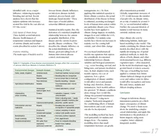
Skin lesions - terms
Type of skin lesion
Raised
Flat
Depressed
Papule
Macule
Erosion
Plaque
Patch
Ulcer
Nodule
Erythema
Atrophy
Cyst
petechiae
Striae
Wheal
Burrow
Scar
Horn
Other characteristics
Shape
round, oval, annular, reticulated, linear, serpiginous, umbilicated
Color of skin lesion
white (leukoderma, hypomelanosis), red/pink (erythematous), violaceous (vascular, ecchymotic)
Consistency
soft, firm, fluctuant
Arrangement
Grouped
herpetiform/zosteriform, arciform, linear, serpiginous,
Disseminated
scattered discrete lesions or diffuse involvement
Distribution
Extent
isolated, localized, generalized
Pattern
symmetrical, exposed areas,
intertriginous
, including palms/solesSlide4
http://medaddicts.blogspot.com/2012/12/terms-used-to-describe-skin-lesions.htmlSlide5
Classification/type of lesion
Raised
Flat
Depressed
Surface Change
Fluid-Filled
Vascular
Papule
Macule
Erosion
Scale
Vesicle
Petechiae
Plaque
Patch
Ulcer
Crust
Pustule
Purpura
Nodule
Erythema
Atrophy
Excoriation
Bulla
Telangiectasia
Cyst
Striae
Fissure
Furuncle
Infarct
Wheal
Burrow
Eschar
Abscess
Scar
Lichenification
Horn
Slide6
Approach
Initial clinical impression (by history/systemic examination)
Classification/type of
lesion
Shape (& color) of individual lesions
Consistency and feel of lesion
Arrangement of multiple lesions
Distribution of multiple lesions
Anatomic components of the skin primarily affected
Approach to Dermatologic DiagnosisSlide7
Fitzpatrick's Dermatology in General Medicine, 7th Edition
papule
nodule
ulcer
crust
vesicle
pustule
macule
plaque
bullae
Shape (description) of lesionsSlide8
Describe the dermatologic condition
CC: Rash
Plaques
Circular/irregular shape
Pink
Smooth
Randomly dispersed
Superficial
http://www.webmd.com/children/ss/slideshow-common-childhood-skin-problemsSlide9
© 2009 WebMD, LLC. All rights reserved.
Skin StructureSlide10
Superficial primary skin infections
Tinea
corporis
–
Patch, oval
,
pink-red (erythematous), scaling
Cutaneous candidiasis –
Plaque, circular/irregular, erythematous
,
with mild scaling
and erythematous satellite
papulesSlide11
Primary Skin Infection
CellulitisSlide12
ErysipelasSlide13
Streptococcal infections
GAS Scarlet Fever
GAS Toxic shock syndrome
Erythroderma
with desquamationSlide14
MeningococcemiaSlide15
Purpura
Fulminans
Meningococcemia
Capnocytophaga
sepsis
Inherited protein C deficiencySlide16
Varicella Zoster Virus
Primary infection - Chickenpox
Virus
DNA
Member of the Herpes family
VZV
prodrome
:
Primary infection: Fever, malaise, loss of appetite, sore throat
Zoster: localized burning pain
Rash
Primary: Papules->vesicle->crust; begins centrally and moves outward
Zoster:
dermatomal
Chickenpox
ZosterSlide17
Herpes Zoster
Vesicles on erythematous baseSlide18
“Poxes”
Smallpox
Variola
virus; a DNA
orthopoxvirus within the Poxviridae
family
Lesions are all in same stage
Chickenpox
Double-stranded
, linear DNA
herpesvirus
Lesions appear in a variety of stages
VesicleSlide19
Rocky Mountain Spotted FeverSlide20
Rocky Mountain Spotted Fever
R.
rickettsii
infects endothelial cells causing
vasculitis
Rash
T
ypically occurs 2-4 days after fever
Begins ankles, wrists and forearms, palms and soles, then spreads centrally
Evolves to petechial lesions
Organism is not evident on blood smearsSlide21
Ehrlichiosis
Infects monocytes and replicates in
cytoplamic
membrane-bound vacuoles
Rash
Much less common (30-40% HME and 1/40 in HE) appears at
approx
5 days
Can be
maculopapular
, macular or petechialSlide22
“Maculopapular rash”…
Henoch-
Schönlein
purpura
Secondary syphilis
Acute HIV
MeaslesSlide23
Secondary SyphilisSlide24
Parvovirus B19
Erythema Infectiosum or Fifth DiseaseSlide25
Sporotrichosis
Fitzpatrick’s Dermatologic in General Medicine, 7
th
EdSlide26
BlastomycosisSlide27
Histoplasmosis
http://www.mjmsr.net/viewimage.asp?img=MullerJMedSciRes_2015_6_1_72_146470_f2.jpgSlide28
Mycobacterium
marinumSlide29
Disseminated Cryptococcus InfectionSlide30
Primary Skin Disorders
Psoriasis
Eczema
https://www.aad.org/public/diseases/scaly-skin/psoriasis
http://www.oregonmedicalresearch.com/patients/active-studies/atopic-dermatitis/Slide31
Rheumatic disease/Autoimmune disease
Systemic Lupus erythematosus
IgA vasculitisSlide32
Cancer-related
Dermatomyositis
Tripe Palm
Heliotrope rash
Gottron’s
papules
https://www.uptodate.com/contents/image?imageKey=DERM%2F61955&topicKey=DERM%2F7606&rank=1~150&source=see_link&search=paraneoplastic%20skin
Acanthosis
nigricansSlide33
Want to learn more about dermatology?
Fitzpatricks
Color Atlas and Synopsis of Clinical Dermatology,
Eighth Edition
/ Edition
8byKlaus Wolff
, Richard Johnson