
Afreen Hasan Electrophoresis is the movement of charged particles through a porous supporting media when subjected to an electric Field Cations move towards cathode Anions move towards anode ID: 908060
Download Presentation The PPT/PDF document "ELECTROPHORESIS By Dr" is the property of its rightful owner. Permission is granted to download and print the materials on this web site for personal, non-commercial use only, and to display it on your personal computer provided you do not modify the materials and that you retain all copyright notices contained in the materials. By downloading content from our website, you accept the terms of this agreement.


Slide1
ELECTROPHORESIS
By
Dr
Afreen
Hasan
Slide2Electrophoresis is the movement of charged particles through a porous supporting media when subjected to an electric Field
• Cations move towards cathode
• Anions move towards anode
• By this technique solutes are separated by their different rates of travel through an electric field which depend on their mass and net electric charge on them
• Commonly used in characterization and differentiation of biological molecules , particularly in the separations of proteins, peptides and nucleic acids
A good clinical example is separation of
isoenzymes
Slide3Factors affecting Electrophoresis
The rate of migration of a solute in an electric field depends on the following factors
Net charge on the particle
2) Mass and shape of the particles
3) p H of the medium
4) Strength of electric field
5) Properties of supporting medium
6) Temperature
Slide4Electrophoretic Mobility
Electrophoretic mobility is defined as the rate of migration (cm/sec) per unit field strength(Volts/cm)
• µ=Q/6π
rη
• Where µ- Electrophoretic mobility
Q-Net charge on the ion r- Ionic radius of the solute
η- Viscosity of the medium
Slide5Electrophoretic mobility
cont
….
The Electrophoretic mobility is directly proportional to net charge and inversely proportional to molecular size/mass and viscosity of the electrophoresis medium
• The p H of solution affects the mobility of the ion by determining the amount and nature of charge
Slide6Electrophoretic mobility
cont
….
•
Proteins, nucleic acids, nucleotides and amino acids bear charged polar groups making them suitable groups for electrophoresis
• Carbohydrates carrying no charged groups are first bound to charged groups like Borate or Sulfite ions and then electrophoresis is carried out
• Lipids are not electrophoresed because electrophoretic current requires polar solvents in which most lipids are insoluble
Slide7Types of Electrophoresis
On the basis of pattern
1) Horizontal
2) Vertical electrophoresis
( mainly used for Polyacrylamide gel electrophoresis
)
Slide8Types of Electrophoresis
cont
…
On the basis of support media on which electrophoresis is performed
(under the heading of zone electrophoresis )
Filter Paper
2) Cellulose acetate membrane (CAM)
3) Agar or Agarose gel
4) Starch Gel
5) Polyacrylamide gel
Some modification of the basic electrophoresis –
Immunoelectrophoresis
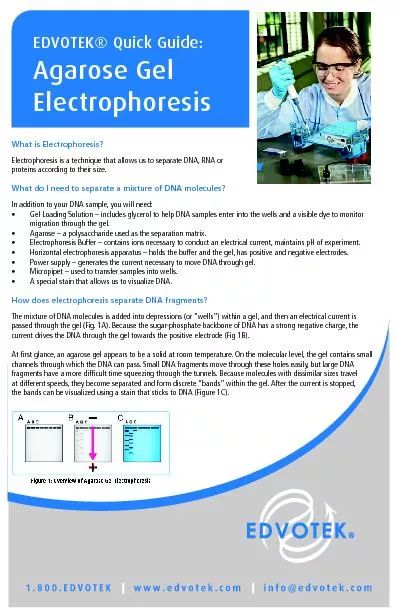
Slide9Slide10Electrophoresis Apparatus
Electrophoresis apparatus consists of
1) Buffer tank -to hold the buffer
2) Buffer
3) Electrodes- made of platinum or carbon
4) Power supply
5) Support media Note-Choice of buffer depends on the nature of substance to be separated and the electricity is supplied at a constant current and voltage.
Slide11Support media for electrophoresis on which electrophoresis can be performed
Filter Paper
2) Cellulose acetate membrane
3) Agar or Agarose gel
4) Starch Gel
5) Polyacrylamide gel
Slide12Paper Electrophoresis
The support medium is a filter paper
• Most common type of electrophoresis run in clinical lab
10µl serum is applied in the form of thin line on hydrated
Whatman
no1 filter paper
Chamber are filled with .1 M
Barbitone
buffer (pH 8.6) and constant current of 1-2 mA
Time duration to run the test is 10-16
hrs
Paper is then stained with Bromophenol blue to visualize individual bands
Drawback- long time interval and blurring of margins
Slide13Slide14Cellulose Acetate Membrane/CAM Electrophoresis
Preferred solid support media
• Less time consuming
• Excellent separation
• No blurring of margins
• Membranes can be stored for a longer time
•
Widely used for separation of
lipo
proteins,
isoenzymes
and hemoglobin variants
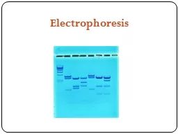
Slide15Gel Electrophoresis
“
Gel" is the matrix used to contain, and then separate the target molecules
• The gel is a cross linked polymer whose composition and porosity is chosen based on the specific weight and composition of material to be analyzed
• A gel block made of Polyacrylamide, Agarose or substituted starch gel is used in this method as the solid support
• Agar gel is used for separation of different types of protein mixtures as well as nucleic acids
•
Polyacrylamide is best suitable for separation of nucleic acids.
It is also frequently used in separating proteins, peptides and amino acids from microgram quantities of mixed samples
Slide16Poly Acrylamide Gel Electrophoresis (PAGE)
Most popular type
• Polyacrylamide is a polymer formed when acrylamide is heated with a variety of catalysts with or without cross linking agents
• It is thermostable, transparent, strong and relatively chemically inert
• Gels are uncharged and are prepared in a variety of pore sizes
•
Proteins are separated on the basis of charge to mass ratio and molecular size, a phenomenon called Molecular sieving.
PAGE is the backbone of blotting techniques
Slide17Types of PAGE
PAGE can be classified according the separation conditions into:
Native-PAGE:
–
Separation is based upon charge, size, and shape of macromolecules.
Useful for separation and/or purification of mixture of proteins – This was the original mode of electrophoresis.
2)
Denatured-PAGE or SDS-PAGE
– Separation is based upon the molecular weight of proteins
. The most common method for determining MW of proteins – Very useful for checking purity of protein samples
Slide18PAGE-Procedure
The gel of different pore sizes is cast in to a column inside a vertical tube, often with large pore gel at the top and small pore gel at the bottom
• Microgram quantity of the sample is placed over the top of the gel column and covered by a buffer solution having such a p H so as to change sample components in to anions
• The foot of the gel column is made to dip in the same buffer in the bottom reservoir
Slide19PAGE PROCEDURE
Slide20PAGE-Procedure
cont
…
Cathode and anode are kept above and below the column to impose an electric field through the column
• Macromolecular anions move towards the anode down the gel column • There is no external solvent space, all the migratory particles have to pass through the gel pores
• Rate of migration depends on the charge to mass ratio
Slide21Visualization of
analytes
on the column of PAGE
After the electrophoresis is complete, the molecules in the gel can be stained to make them visible.
• Ethidium bromide, silver, or
comassie
blue dye may be used for this process.
• If the
analyte
molecules fluoresce under ultraviolet light, a photograph can be taken of the gel under ultraviolet lighting conditions.
If the molecules to be separated contain radioactivity added for visibility, an autoradiogram can be recorded of the gel.
Slide22SDS PAGE
SDS-PAGE, sodium dodecyl sulfate polyacrylamide gel electrophoresis, is a technique widely used in biochemistry, forensics, genetics and molecular biology to separate proteins according to their electrophoretic mobility
• The SDS gel electrophoresis of samples having identical charge to mass ratios results in fractionation by size
Probably the world's most widely used biochemical method
Slide23SDS PAGE
cont
….
When a detergent SDS(Sodium -
DodecylSulfate
)is added to PAGE the combined procedure is termed as SDS PAGE
• SDS coats protein molecules giving all proteins a constant charge-mass ratio.
• Due to masking of charges of proteins by the large negative charge on SDS binding with them, the proteins migrate along the gel in order of increasing sizes or molecular weights
Slide24SDS PAGE
cont
…..Molecular weight of a given protein can be determined by comparing the relative electrophoretic mobility of sample with that of standard protein of known molecular weight, when both the sample and the standard proteins are electrophoresed side by side in the same gel slab
• In
oligomeric
proteins, SDS PAGE usually gives the molecular weight of separated monomer chains of the proteins because SDS cleaves the non covalent bonds interlinking the monomer chains in the intact molecule.
Slide25SDS PAGE
Slide26ISOELECTRIC FOCUSSING
Every protein has its own isoelectric point
pI
ie
pH at which they are neutral in electric field
Gels are impregnated with
ampholine
Ampholine
makes a continuous pH gradient in Polyacrylamide gels
Loading of sample /protein on these
imprignated
gel and kept in electrical field cause focusing of proteins to their
pI
Formation of sharp bands occurs
Eg
–separation of different
isoenzymes
clinically
Slide27IMMUNOELECTROPHORESIS
Antigens and antibodies are allowed to interact with each other in agarose gel
Old one immune electrophoresis called as Rocket electrophoresis
Antibodies are embedded in agarose gel at the time of its solidification
Antigen mixture is allowed to migrate into the gel by electrophoresis
Frequently done in clinical serology
Slide28IMMUNOELECTROPHORESIS
cont
….
Crossed
immunoelectrophoresis
–(a variety of
immunoelectrophoresis
)
Both antigen and antibodies move towards each other under the influence of electrical field and form precipitation lines at places where the concentration of both are
equimolar
Slide29IMMUNOELECTROPHORESIS
cont
….
Immunofixation
– (a variety of
immunoelectrophoresis
)
Proteins are run on a routine electrophoresis and then exposed to specific antibodies
Antibodies act as probes for identification of antigens present in low concentration and make precipitation line
Unreacted proteins are washed
Precipitin lines are visualized with certain stains
Slide30Slide31