
SYSTEMIC HISTOLOGY II ATIBA PM OVARY MESOTHELIAL EPITHELIUM Simple cuboidal or Columnar TUNICA ALBUGENIA Dense connective tissue Histology of Female Reproductive System 2 OVARY CORTEX ID: 917159
Download Presentation The PPT/PDF document "HISTOLOGY OF FEMALE REPRODUCTIVE SYSTEM" is the property of its rightful owner. Permission is granted to download and print the materials on this web site for personal, non-commercial use only, and to display it on your personal computer provided you do not modify the materials and that you retain all copyright notices contained in the materials. By downloading content from our website, you accept the terms of this agreement.


Slide1
HISTOLOGY OF FEMALE REPRODUCTIVE SYSTEM
SYSTEMIC HISTOLOGY
II
ATIBA, P.M
Slide2OVARY
MESOTHELIAL EPITHELIUM
Simple cuboidal or Columnar
TUNICA ALBUGENIA
Dense connective tissue
Histology of Female Reproductive System
2
Slide3OVARY
CORTEX
Stroma houses ovarian follicles at various stages of development
MEDULLA
Loose connective tissue containing vascular bed and nervous
Histology of Female Reproductive System
3
Slide4OVARIAN CYCLE
Three Phase of ovarian cycle:
Follicular Phase
Development of primordial follicular-Mature follicleFollicular phase of endometriumOvulatory PhaseRelease of oocyte from mature Follicle and received by fimbrae of oviductLuteal PhaseResidual follicular cell folds and becomes part of Corpus Luteum (C.L)Secretion/luteal phase of endometriumHistology of Female Reproductive System
4
Slide5FOLLICULAR GROWTH
Ovarian follicle is;
An oocyte
Follicular / granulosa cells
PRIMORDIAL FOLLICLE
(formed during fetal life)
Follicular Growth
Modification of:
Oocyte
Granulosa cells
Stromal Fibroblast
Histology of Female Reproductive System
5
Slide6FOLLICULAR GROWTH (Follicular Phase)
Histology of Female Reproductive System
6
Slide7PRIMORDIAL FOLLICLE :
Primary Oocyte
Arrested in Prophase stage of Meiosis 1
about 25µm in diameterFollicular cellsSingle layer of flattened cells
Attached by desmosome
FOLLICULAR GROWTH (Follicular Phase)
Histology of Female Reproductive System
7
Slide8FOLLICULAR PHASE
PRIMARY FOLLICLE
Primary oocyte
Grows to 125-150µm diameter
Follicular cellsCuboidal cells
More than 1 layer
Zona
pellucida
separate oocyte from Follicular cells
Histology of Female Reproductive System
8
Slide9FOLLICULAR PHASE
SECONDARY FOLLICLE
Primary oocyte
Grows to 125-150µm diameter
Follicular cells
Cuboidal cellsCumulus
oophorus
Stromal cells
Theca
interna
(steroid producing)
Theca externa
Histology of Female Reproductive System
9
Slide10GRAAFIAN FOLLICLE
Primary oocyte
Follicular cells
Increases to about 2.5cm in diameter
Increase in size follicular antrumThe oocyte become eccentrically located
FOLLICULAR PHASE
Histology of Female Reproductive System
10
Slide11Day 14 of menstrual cycle
LH surge
Rupture wall of
Graafian
folliclePG, Histamine, Collagenases, Hyaluronic acidRelease of secondary oocyte (arrest in metaphase II) with corona radiate
Oocyte received by fimbrae
OVULATION PHASE
Histology of Female Reproductive System
11
Slide12AFTER OVULATION
Remnant of
graafian
follicle collapse and folded
Blood flow to follicular cavity and innveded by Connective tissue- phagocytes in central part of corpus luteumGranulosa cell- granulosa lutein cellsTheca interna cells- theca lutein cells
Oestrogen and Progesterone produced by Corpus luteum
LUTEAL PHASE
Histology of Female Reproductive System
12
Slide13DEPENDENT ON ESTABLISHMENT OF PREGRNANCY OR NOT?
If it occurs Corpus luteum degenerate- corpus
albicans
hCG
maintains C.L – C.L of pregnancy- secrete hormone – maintain pregnancyFATE OF CORPUS LUTEUMHistology of Female Reproductive System
13
Slide14C.L of menstruation is invaded by
fibrblasts
– fibrotic
Remnant undergoes
luteolysisFibrous connective tissue – corpus albicansPersist as the scar on the surface of ovary
CORPUS ALBICANS
Histology of Female Reproductive System
14
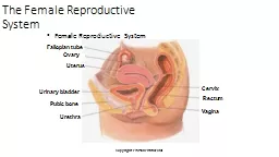
Slide15The walls composed of:
Mucosa layer
Lines by simple columnar epithelium
Lamina
propriaCharacterised by longitudinal foldsMuscularis
LayerInner circular and outer longitudinal layers of smooth muscle
Serosa layer
Simple squamous epithelium
UTERINE TUBE
Histology of Female Reproductive System
15
Slide16Two types epithelium cell:
Non ciliated pigment cells
No cilia
Secretory:
Nutritive and protective for oocyteCapacitation of spermCiliated cellsCilia beat toward the uterus
UTERINE TUBE
Histology of Female Reproductive System
16
Slide17UTERUS
The walls of Uterus:
Endometerium
Myometerium
Serosa/Adventitia
Histology of Female Reproductive System
17
Slide18ENDOMETRIUM
Epithelium
Simple columnar epithelium
Ciliated cells
Secretory cells
Lamina Propria
Loose Connective tissue, rich in
firoblast
, reticular
fibre
Uterine glands (simple tubular)
Undergoes cyclic changes
Histology of Female Reproductive System
18
Slide19Two Layers (Zone)
Functionalis
Thick, superficial (surface epithelium, lamina
propria
and gland)Rich capillary network (coiled arteries)Sloughed at menstruationBasalis
Deep, narrow (lamina propria
)
Straight arteries
Regenerate
functionalis
layer each menstrual cycle
ENDOMETRIUM
Histology of Female Reproductive System
19
Slide20Myometrium
Thickest layer of Uterus
Composed of three layers of smooth muscle:
Inner longitudinal layer
Middle circular
Outer longitudinal layerThe size and number of muscle cells are related to estrogen levels
Pregnancy: hyperplasia and hypertrophy
Histology of Female Reproductive System
20
Slide21Under stimulus of estrogen and progesterone endometrium undergoes cyclic structure modification
Duration: 28days
Puberty Menopause
Menstrual cycle
Histology of Female Reproductive System
21
Slide22Proliferative/follicular Phase (Days 5-14)
After menstrual phase
Coincides with ovarian follicles development (estrogen)
Regeneration of endometrium
Surface epithelium
Lamina propria
Coiled arteries
Uterine glands: simple columnar epithelial, straight tubule, narrow lumens
Histology of Female Reproductive System
22
Slide23Secretory/Luteal Phase (days 15-28)
Begins after ovulation
Depends on corpus luteum secretion (progesterone)
Uterine glands: coiled and branched, accumulation of glycogen dilate the lumen
Thickening of
functionalisPrepared to receive zygote
Histology of Female Reproductive System
23
Slide24Menstrual Phase (days 1-4)
No fertilization corpus luteum degenerates(result in decrease in progesterone and estrogen)
Coiled arteries constrict
functionalis
layer becomes ischemic necrotic Rupture of arteries hemorrhagic
Functional layer shed off
Basal layer restore functional layer
Histology of Female Reproductive System
24
Slide25ENDOMETRIAL CYCLE
Histology of Female Reproductive System
25
Slide26PLACENTA
Transient organ- site of physiologic exchange between mother and fetus.
Endocrine function
Consist of;
Fetal part
Chorionic villi arise from chorionic plate
Connective tissue surrounded by
syncytiotrophoblast
and
ctyotrophoblast
Mother part
Decidua basalis from lacunae
Histology of Female Reproductive System
26
Slide27PLACENTA
Placenta barrier
Trophoblast layers
Basal lamina of trophoblast
Mesenchyme
Basal lamina of capillaries
Endothelium of fetal capillary
Histology of Female Reproductive System
27
Slide28CERVIX
Epithelium
Lumen; mucus –secreting simple columnar epithelium
External surface; stratified squamous non-keratinized epithelium
WallDense, collagenous connective tissue
Cervical glands regulates by progesterone
Serous /watery fluid; around ovulation time
Viscous/mucus; at pregnancy/luteal phase of menstruation
Histology of Female Reproductive System
28
Slide29VAGINA
It consists of three layers:
Mucosa
Stratified squamous on keratinized epithelium
Lamina propria
; loose fibroelastic connective tissue, rich vascular
No glands; vaginal fluid comes from transudation and cervical glands
Muscularis
Smooth muscle, inner circular and outer longitudinal
Adventitia
Dense
fibroelastic
connective tissue
Histology of Female Reproductive System
29
Slide30EXTERNAL GENITALIA
Labia
Majora
Skin
Rich adipose tissueSweat and sebaceous glands
Labia Minora
Spongy connective tissue with elastic
fibre
Less blood vessel and nerve ending
Sebaceous glands
Histology of Female Reproductive System
30
Slide31EXTERNAL GENITALIA
Clitoris
Stratified squamous epithelium
2 erectile bodies (blood vessels, sensory nerve)
GlandsGlands of Bartholin
Minor vestibular glands
Histology of Female Reproductive System
31
Slide32MAMMARY GLANDS
Organs of milk production
Consist of 15-25 lobes of compound
tubuloaveolar
gland
Excretory lactiferous duct
Dense connective tissue separate the lobes
Variation in structure:
Age
Physiologic status
Histology of Female Reproductive System
32
Slide33ENDOMETRIAL CYCLE
Histology of Female Reproductive System
33