
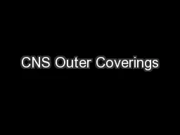
Brain cranial bones Spinal Cord vertebrate Inner Coverings Meninges Dura mater Arachnoid membrane Pia mater Pia Mater Arachnoid Membrane Dura Mater Epidural Space Subdural Space Subarachnoid Space ID: 572871
Download Presentation The PPT/PDF document "CNS Outer Coverings" is the property of its rightful owner. Permission is granted to download and print the materials on this web site for personal, non-commercial use only, and to display it on your personal computer provided you do not modify the materials and that you retain all copyright notices contained in the materials. By downloading content from our website, you accept the terms of this agreement.


Slide1Slide2Slide3Slide4Slide5
CNS
Outer Coverings
Brain
cranial bones
Spinal Cord
vertebrate
Inner Coverings
Meninges
Dura mater
Arachnoid membrane
Pia materSlide6
Pia Mater
Arachnoid Membrane
Dura Mater
Epidural Space
Subdural Space
Subarachnoid Space
sc
vertebrate
Pia
Arachnoid
Dura
Subarachnoid Space (contains CSF)
Subdural Space
Epidural SpaceSlide7
Meninges
Dura Mater
Outer layer
Strong, white fibrous
tissue
Falx
cerebri
– extension
of dura
mater that
extends vertically
to separate two hemispheresArachnoid MembraneMiddle layerDelicate, cobwebby Pia MaterInner layer (adheres to outer surface of brain and spinal cord)TransparentContains blood vesselsSlide8
Meninges Spaces
Epidural Space
Between dura mater and bony covering of brain and spinal cord
Supportive cushion of fat
Subdural Space
Between dura mater and arachnoid membrane
Lubricating serous fluid
Subarachnoid Space
Between arachnoid and pia mater
Contains cerebrospinal fluid (CSF)Slide9Slide10Slide11
Meningitis
infection/swelling of meninges
caused by infection with viruses, bacteria, or other microorganisms
may also arise due to certain drugs or other diseases.
potentially life threatening due to the inflammation's proximity to the brain and spinal cord; it is therefore a medical emergency
symptoms
headache and neck stiffness
Fever, confusion or altered consciousness
inability to tolerate light (photophobia) or loud noises (phonophobia).
Sometimes, especially in small children, only nonspecific symptoms may be present, such as irritability and drowsiness.
If a rash is present, it may indicate a particular cause of meningitis (meningococcal bacteria
diagnosed by a spinal tap
must be treated promptly with antibiotics and sometimes antiviral drugs In some situations, corticosteroid drugs can also be used to prevent complications from overactive inflammation. can lead to serious long-term consequences such as deafness, epilepsy, hydrocephalus and cognitive deficit, especially if not treated quickly. Some forms of meningitis may be prevented by immunizationSlide12
CSF
Provides supportive, protective cushioning
Reservoir of circulating fluid
Monitored by brain to detect changes in internal environment
Located in subarachnoid space and within cavities and canals of brain and spinal cord
Average adult has 140ml of CSFSlide13
Hydrocephalus
“water head”
Sometimes in the unborn child, the drainage canal for CSF becomes stopped up.
The fluid builds up and the pressure causes the brain to expand like a balloon.
Causes the child to have a very large head and to be mentally retarded
Accompanies diseases (spina bifida, brain tumor, blood clots)
Possible coma or deathSlide14Slide15
Spinal Cord
Within spinal cavity (vertebral column)
Extends from foramen magnum to L
1
Reflex center
Dorsal nerve root
carries sensory info
into
spinal cord
Ventral nerve root
carries motor info
out of spinal cordInterneurons – in s.c. gray matterSpinal nerve – single mixed nerve on each side of s.c where dorsal and ventral nerve roots joinSlide16
Spinal Cord
Gray Matter
Extends length of s.c
Consists of cell bodies of interneurons and motor neurons
Spinal reflex centers located here
Incoming sensory, outgoing motor
White Matter
Surrounds gray matter
Consists of axonsSlide17
Spinal CordSlide18
Brain
One of largest organs in adults
3 lbs
6 major divisions
Medulla oblongata
Pons Brainstem
Midbrain
Cerebellum
Diencephalon
CerebrumSlide19
Internal (Half) Brain
Optic chiasmSlide20
Brainstem
Medulla Oblongata
Lowest part of brainstem
Attaches brain to
s.c.
just above foramen magnum
Reticular Formation – arousal, sleep (damaged=coma) [
R
eticular
A
ctivating
System]Controls breathing, heart rate and the activities of the gut Coordinates swallowing, yawning, hiccuping, vomiting, coughing and sneezing
Injury often causes death https://www.youtube.com/watch?v=cu7A8LIzL1oSlide21
Brainstem
Pons
Between medulla and midbrain
motor control and sensory analysis
Regulate respirationSlide22
Brainstem
Midbrain
Above pons, below cerebrum
Auditory and visual centers
Muscular controlSlide23
Cerebellum
2
nd
largest part of brain
Numerous sulci (grooves) and gyri (raised area)
Acts with cerebral cortex to produce skilled movements (coordination)
Controls skeletal muscles for balance
Controls posture
Subconscious level; automatic processor
Impulses travel from cerebellum to cerebrum and muscles to coordinate movementSlide24
Diencephalon
Between cerebrum and midbrain
Consists of
Thalamus
Hypothalamus
Optic chiasma
Pineal bodySlide25
Diencephalon
Thalamus
Major relay station for sensory impulses on their way to cerebral cortex
Sensations
Conscious recognition of pain, temperature, touch
Relay sensory info (except smell) to cerebrum
Emotions of pleasant and unpleasantness
Complex reflexesSlide26
Diencephalon
Hypothalamus
Below thalamus
Links mind and body
Regulates and coordinates autonomic activities
Synthesizes hormones secreted by pituitary gland
Water balance
Regulates appetite
Maintains normal body
temperatureSlide27
Diencephalon
Pineal Body
Regulates body’s biological clock
Produces some hormones
MelatoninSlide28
Cerebrum
Cerebral cortex, cerebral tracts, cerebral nuclei.
Four general functions
Consciousness
Language
Emotions
Memory
Gyri (bumps) and sulci (shallow grooves)
Fissures – deep grooves, divides lobes
Longitudinal fissure – divides hemispheres
Central sulcus – between frontal and parietal lobes
Lateral fissure – between temporal and parietal lobes
Parietooccipital fissure – between occipital and parietal lobesOuter surface made up of 6 layers of gray matterLargest and uppermost division of brainRight and left hemispheres Separated by corpus collosum
Each hemisphere has 4 lobesFrontal ParietalTemporal occipital
Parietooccipital fissure
Lateral fissureSlide29
Frontal lobe
Prefrontal:
Personality
And adaptation of the personality to events and experiences
Foresight and imagination
Sense of self
Frontal:
main motor areas (originate movement that is coordinated elsewhere)
Broca’s Area:
speech productionSlide30
Parietal lobe
Principle sensory area
Touch
Proprioception
Lesions cause sensory losses
Involvement in cognition
Receptive speech lossSlide31
Temporal lobe
Cognition
Emotion
Memory
Auditory
Wernicke’s area:
speech comprehensionSlide32
Occipital lobe
Vision
Visual processing and visual association
Involved in eye movementSlide33
Limbic System
emotion, behavior,
long term memory, and olfaction
Set of brain structures that forms the inner border of the cortex
Corpus callosum:
connects left and right hemispheres
Hippocampus:
long-term memory; cognitive maps
Amygdala:
reward, fear, matingSlide34
Left Hemisphere
Language
Dominating hand movements
Reasoning (tangible data)
Positive emotion
Right Hemisphere
Hearing
Touch
Spatial relationships
Nonsymbolic data
Art
Spiritual
Negative emotionsSlide35Slide36Slide37Slide38
busy wave
relaxed wave
drowsy wave
deep sleep wave
EEG/ECG
ElectroencephalogramSlide39Slide40
CNS Disorders
Aphasia
loss of speech
https://
www.youtube.com/watch?v=1aplTvEQ6ew
Hemiplegia, paraplegia,
triplegia
, quadriplegia
paralysis
Cerebral palsy
crippling
disease involving permanent damage to motor control areas of the brainSpastic paralysisaltered skeletal muscle performance in muscle tone involving hypertonia; it is also referred to as an unusual "tightness", stiffness, or "pull" of muscles
lack of inhibition results in excessive contraction of the muscles, ultimately leading to hyperflexia (overly flexed joints)Presents in multiple sclerosis and other CNS disordersCVA (cerebrovascular accident) aka Stroke cessation or hemorrhage of blood causing neuronal damagehttps://www.youtube.com/watch?v=6KpYjUJ2t0oDementiaAlzheimer’s: inherited form of dementia (early signs around age 30-40)
https://www.youtube.com/watch?v=cqmZFoGvzfUHuntington’s Disease: affects memory in middle to late adulthood, causing cortex lesionsAIDSSeizuresEpilepsy https://www.youtube.com/watch?v=6NcqQkKjqTI
https://www.youtube.com/watch?v=YDA7gGjpPWMSlide41Slide42
Somatic Nervous System
Contraction of skeletal muscles
Skeletal muscle = somatic effector
All
voluntary
motor pathways
outside
of CNS
Neurotransmitter = AChSlide43
Reflexes
All voluntary motor pathways outside of CNS
Reflexes
Action resulting from nerve impulse passing over a reflex arc
Predictable response to stimuli
Autonomic Reflex
Visceral
Contraction of smooth or cardiac muscle
Secretion of glands
Somatic Reflex
Contraction of skeletal musclesSlide44
Somatic Reflexes
Contraction of skeletal muscles
Reflexes deviate from normal in certain conditions
Reflex testing is valuable diagnostic tool
Patellar Reflex: extension of lower leg
Achilles Reflex: extension of foot
Babinski Reflex: extension of big toe
Present until age 1.5
If present after, indicates damage to corticospinal fibers
Plantar Reflex: flexion of all toes and slight turning in of foot
Corneal Reflex: wink when touch cornea
Abdominal Reflex: stroke side of abdomen causes drawing in of abdominal wallSlide45
Knee-Jerk (Patellar) ReflexSlide46
Autonomic Nervous System
Involuntary/Visceral body functions
Cardio, resp, dig, urogen
Maintain homeostasis by: regulating heartbeat, smooth muscle contraction, glandular secretions
Conduct impulses
from
CNS to autonomic effectors
Two divisions
Sympathetic
ParasympatheticSlide47
Autonomic Conduction PathwaySlide48
Parasympathetic Nervous System
“Feed-or-Breed”
“Rest-and-Repose”
Counteracts Sympathetic
“Fight-or-Flight”
Allows body to function under stress
Sympathetic
Nervous SystemSlide49Slide50
ANS Neurotransmitters
Norepinephrine (adrenaline)
Adrenergic fibers
release
norepinephrine
in
post
synaptic
sympathetic
neurons
Acetylcholine (ACh)
Cholinergic fibers release ACh in presynaptic sympathetic neuronsrelease ACh in pre and post parasympathetic neuronsSlide51Slide52
Norepinephrine
Affects visceral effectors by binding to adrenergic receptors
Alpha receptor: blood vessels constrict
Beta receptor: blood vessels dilate
Inhibiting action of norepinephrine
MAO (monoamine oxidase): enzyme that breaks up norepinephrine that is taken up by synaptic knobsSlide53Slide54
ACh
Binds to cholinergic receptors
Nicotinic receptors
Muscarinic receptors
Inhibiting action of Ach
acetylcholinesteraseSlide55