
SYMPTOMSLUMPS Professor P Grantley Gill Specialists Without Borders Seminar in Surgery Rwanda September 2010 SIGNS AND SYMPTOMS AT PRESENTATION Palpable mass Thickening Pain Mass or pain in the axilla ID: 335280
Download Presentation The PPT/PDF document "ASSESSMENT OF BREAST" is the property of its rightful owner. Permission is granted to download and print the materials on this web site for personal, non-commercial use only, and to display it on your personal computer provided you do not modify the materials and that you retain all copyright notices contained in the materials. By downloading content from our website, you accept the terms of this agreement.


Slide1
ASSESSMENT OF BREAST
SYMPTOMS/LUMPS
Professor P Grantley Gill
Specialists Without Borders
Seminar in Surgery
Rwanda, September 2010Slide2
SIGNS AND SYMPTOMS AT PRESENTATION
Palpable mass
Thickening, Pain
Mass or pain in the axilla
Nipple discharge
Oedema or erythema of the skin
www.specialistswithoutborders.orgSlide3
CAUSES OF BREAST LUMPS
Fibroadenoma
CystFibrocystic change
CancerDuct ectasiaFat necrosisHaematoma
www.specialistswithoutborders.orgSlide4
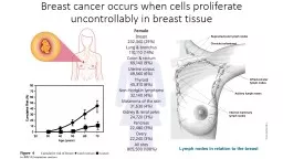
BREAST CANCER RISK FACTORS
Age
History of breast cancer
Family history of breast cancer, especially in first degree relativesSpecific genetic mutations e.g. BRCA-1, BRCA-2
Benign breast “cancer” atypical hyperplasia
Early menarche, late menopause
Late first pregnancy/no pregnancy
Exogenous oestrogensRadiationDiet, alcoholwww.specialistswithoutborders.orgSlide5
EXAMINATION OF BREAST
Inspection
Palpation
Lymph node basins (axilla, neck)Contralateral breast
www.specialistswithoutborders.orgSlide6
INSPECTION
www.specialistswithoutborders.orgSlide7
NIPPLE RETRACTION
www.specialistswithoutborders.orgSlide8
SKIN DIMPLING
www.specialistswithoutborders.orgSlide9
PAGET’S DISEASE
www.specialistswithoutborders.orgSlide10
PAGET’S DISEASE
www.specialistswithoutborders.orgSlide11
TRIPLE ASSESSMENT OF A BREAST LUMP
Clinical
ImagingMammography
UltrasoundPathologyFine needle aspiration cytology (FNAC)Core biopsy
www.specialistswithoutborders.orgSlide12
PALPABLE LYMPH NODES
www.specialistswithoutborders.orgSlide13
MAMMOGRAPHIC STAGING
Masses
Asymmetry
Architectural distortionCalcificationSkin changes
www.specialistswithoutborders.orgSlide14
www.specialistswithoutborders.org
NORMAL FATTY REPLACEMENTSlide15
www.specialistswithoutborders.org
NORMAL INVOLUTION
Slide16
DENSE BREASTS
www.specialistswithoutborders.orgSlide17
DENSE BREASTS – RETROAREOLAR TISSUE LEFT
www.specialistswithoutborders.orgSlide18
DENSE BREASTS +++ ALL OF LIFE
www.specialistswithoutborders.orgSlide19
ULTRASOUND
A: Fibroadenoma
B: Carcinoma
www.specialistswithoutborders.orgSlide20
FINE NEEDLE ASPIRATION CYTOLOGY (FNAC)
www.specialistswithoutborders.orgSlide21
www.specialistswithoutborders.orgSlide22
FNAC INTERPRETATION AND ACTION
www.specialistswithoutborders.org
INTERPRETATON
ACTION
Unsatisfactory for diagnosis
Repeat or core
Cellular, benign
Accept if consistent with imaging; repeat or core if not
Cellular, some atypia
Core biopsy
Open biopsy
Suspicious
Core biopsy
Open biopsy
Malignant
TreatmentSlide23
TRIPLE ASSESSMENT OUTCOME
www.specialistswithoutborders.org
ASSESSMENT
CONCORDANT
DISCORDANT
Clinical
Accept results benign or malignant
Core biopsy
Open biopsy
Imaging
CytologySlide24
CONCLUSION
Confirm malignancy in ≥90% by triple assessment
Open biopsy if not possible
www.specialistswithoutborders.org