

DHO Book p470549 amp Nursing Assistants p315 Terminology Quiz Friday March 15 th Odontology Study of the anatomy growth and diseases of the teeth Crown section of the tooth that is visible in the mouth ID: 278935
Download Presentation The PPT/PDF document "Dental" is the property of its rightful owner. Permission is granted to download and print the materials on this web site for personal, non-commercial use only, and to display it on your personal computer provided you do not modify the materials and that you retain all copyright notices contained in the materials. By downloading content from our website, you accept the terms of this agreement.


Slide1
Dental
DHO Book p.470-549 & Nursing Assistants p.315Slide2
Terminology – Quiz Friday March 15
th
Odontology - Study of the anatomy, growth, and diseases of the teeth
Crown – section of the tooth that is visible in the mouth
Root – section of tooth located below the gingiva or gums
Enamel – hardest tissue in the body and covers the outside of the crown. Made mostly of calcium and phosphorus. It is the protective layer of the tooth. Enamel does not grow or repair itself after it is formed.
Dentin – tissue that makes up the main bulk of the tooth. It is bonelike, but softer than the enamel. Dentin is living tissue and can repair and grow.
Pulp = soft tissue located in the innermost area of the tooth. Mostly made up of blood vessels and nerves and held in place by connective tissue.
Periodontium = structures that surround and support the teeth
8. Gingiva – gums – made of epithelial tissue covered by mucous membrane.Slide3
Terms Cont.
Primary / deciduous teeth – The first set of teeth (20). Often called “baby teeth”
Incisors – teeth located in front of mouth, sharp, used to cut or bite food
Cuspids – aka canines, or eyeteeth – used to tear food, longest teeth in the mouth
Bicuspids – premolars, pulverize or grind food
Molars – teeth located in the back of the mouth – largest and strongest – chew and grind food
Universal Numbering System – abbreviated form for identifying the teeth
Anterior – “towards the front”
Posterior – towards the back
Labial – crown surface next to the libs
Lingual – crown surface next to the tongue
Incisal = cutting edge of the tooth
Mesial – side surface closest to the midlineSlide4
Terms Cont.
Distal – side surface away from the midline
Restoration – the process of replacing a diseased portion of a tooth or a lost tooth by artificial means
Cavity – when enamel, dentin and or cementum are destroyed – creates a hollow space.
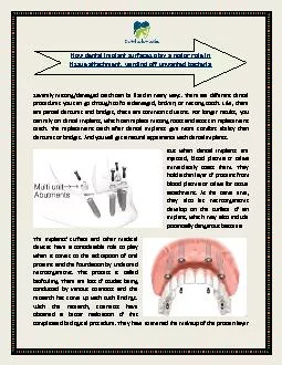
Amalgam – restorative material used primarily on posterior teeth – usually made of a mixture of metals
Composite – restorative material used most frequently in repair of anterior teeth but now being used in posterior as well. Becoming stronger. Applied in layers and uses a curing lightSlide5
The tooth – page 471 Figure 17-2
You will be responsible for knowing all parts of the tooth as illustrated on this page. Be prepared to label a diagram.
Stop: complete activity Slide6
Dental
Odontology is the study of the anatomy, growth, and diseases of the teeth.
Teeth:
1. Accessory organs of the digestive tract
a. Mastication or chewing of food
2. Two dentitions (sets of teeth)
a. Primary dentitions – 20 teeth
i. At birth a newborn has 44 tooth buds
ii. At 6 months teeth begin to errupt
iii. By 2 years – all primary teeth have eruptedSlide7
Teeth Continued
* between ages 6-12 all primary teeth are lost and replaced by permanent
dentition
B. Permanent Teeth – 32 teeth
*begin to errupt at age 5
*continue until 17-20 years old
*most are in place by age 12
Slide8
4 main parts of a tooth
Crown – section of the tooth that is visible in the mouth and protected by enamel
2. Root – below the gingiva or gums, covered by by cementum. It anchors or holds tooth in the bony socket of the jaw
3. Cervix = also called the neck or cemento-enamel junction. It is the area where the enamel covering the crown meets the cementum covering the root.
Apex – Tip of the root of the tooth. Contains an opening called the apical foramen, this is where the blood supply and nerves enter the tooth.Slide9
4 main tissues that make up a tooth
Enamel – hardest tissue in the body, covers the crown, made of mostly calcium and phosphorus and forms a protetive layer for the tooth. It does not grow or repair itself.
Cementum – hard, bonelike tissue that covers the outside of the root. It also provides a thin layer of protection and holds the tooth in place. It forms continuously thoughout life.
Dentin – The main bulk of the tooth, bonelike and softer than the enamel but harder then the cementum. It lacks nerves, but it still carries sensations of pain and temperature to the pulp of the tooth. The Dentin is living tissue, it may repair and grow. The internal surface of the dentin makes the wall of the pulp chamber.
Pulp – Soft tissue / innermost area of the tooth. Made of blood vessels and nerves, held in place with connective tissue.
Pulp chamber – portion of pulp located in crown
Pulp canal or root canal – portion contained in the root
Pulp cavity - the chamber and canal create a space in the tooth – provides nourishment and produces dentin.Slide10
The Periodontium
Structures that surround and support the teeth
Alveolar process or ridge – upper and lower jaw
Periodontal ligament – connective tissue that attaches to the cementum of the tooth and to the alvelous. It supports the tooth in the socket and absorbes shock during chewing. It contains nerves and blood to nourish the tooth, aides in the production of cementum and produces sensation when pressure is applied.
Gingiva – aka Gums – made of epithelial tissue covered with mucous membrane. Covers the alveolar bone and surrounds the teeth. Slide11
Stop Day 2 notes
Answer Questions 3 & 4 on page 473 of DHO book (under procedure). For # 4 use the diagram passed out on Monday.Slide12
Identifying the teeth
Incisors
located in the front and center of the mouth
broad, sharp edge
used to cut or bite food
central incisors are in the center
lateral incisors are on the sides of the centralsSlide13
Cuspids
also known as canines, or eyeteeth
located at angles of lips
used to tear food
longest teeth in the mouthSlide14
Bicuspids
also known as premolars
located before the molars, from front to back
used to pulverize or grind food
*no bicuspids in primary dentitionSlide15
Molars
Teeth in the back of the mouth
Largest and strongest teeth
Used to chew and grind foodSlide16
Identifying Teeth
Four Quadrants
1. Maxillary Right
2. Maxillary Left
3. Mandibular Right
4. Mandibular LeftSlide17
Universal Numbering System
An abbreviated form for identifying the teeth. Used by the American Dental Association and is the main way to identify teeth.
Primary teeth labeled A-T
Starts at Maxillary right, then maxillary left, mandibular left, and finishes mandibular right
Permanent teeth – numbered 1-32 using the same quadrants as above.Slide18
Stop Day 3 notes
Complete questions 5-7 on page 473 of DHO bookSlide19
Careers in the Dental Field
Dentist
Dental Assistant
Dental Hygienist
Orthodontist, Periodontist, Endodontist, Oral Surgeon
Prostetics
Watch the video of the dental assistant and hygienist – write a 1-2 paragraph summary of what you learned.
Link to video:
http://wps.prenhall.com/phschool_colbert_anat_pathophsy_1/99/25575/6547234.cw/index.htmlSlide20
Care of patient’s teeth (in clinical setting)
Mosby’s textbook for Nursing Assistants
Chapter 16
Review Assisting the person to brush the teeth p. 316
Review Brushing the Person’s Teeth p.317
Review Flossing the Person’s teeth p 318 -319
Review Mouth Care for the Unconscous Person p 320-321
Review Denture Care p.322-323
We will be practicing all of these skills in class / you must participate fully in order to receive credit.